

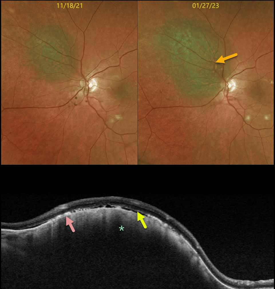
This 71YO female presented on 1/27/23 for follow-up for an asymptomatic, less than 1mm thick choroidal nevus measuring 6.3 x 5.5 mm.
Optos color imaging shows that the nevus has significantly increased in size (3.0 x 10.5 x 7.5 mm) compared with the prior visit on 11/18/21. The nevus enlarged laterally in all dimensions and now touched the nerve margin. There was significantly more overlying orange pigment.
Swept-source OCT shows a thickened choroidal lesion. The clinically noted orange pigment is seen on OCT as hyperreflective outer retinal deposits. Trace variable subretinal fluid vs loss of outer retinal tissue is also seen.
Radioactive plaque placement was performed. Fine needle biopsy revealed the tumor to be Class 1A, PRAME negative.
Learning Points:
Clinical characteristics of choroidal nevi at risk for progressing to a uveal MM were initially described by Shields et al (Ophthalmology 1995;102:1351-1361). The most recent version of this famous acronym (Shields et al, Retina 2019;39:1840-1851) is To Find Small Ocular Melanoma Doing Imaging (TFSOM-DIM), which stands for:
To (Thickness >2mm)
Find (subretinal Fluid)
Small (Symptoms, vision <= 20/50)
Ocular (Orange pigment)
Melanoma (Melanoma hollow on ultrasonography)
DIM (DIaMeter > 5mm).
Although our patient had several of these baseline risk factors (orange pigment, 6.3mm diameter), the growth in just 6 months is unusual.
Patients with Class 1A lesions have a lower 5-year metastatic risk (2%) than those with Class 1B or 2 lesions. PReferentially expressed Antigen in MElanoma (PRAME) is a gene that can be aberrantly expressed in some malignancies. Its positivity can indicate an increased risk for metastatic disease in Class 1 tumors, but its negativity does not tend to alter prognosis.



