

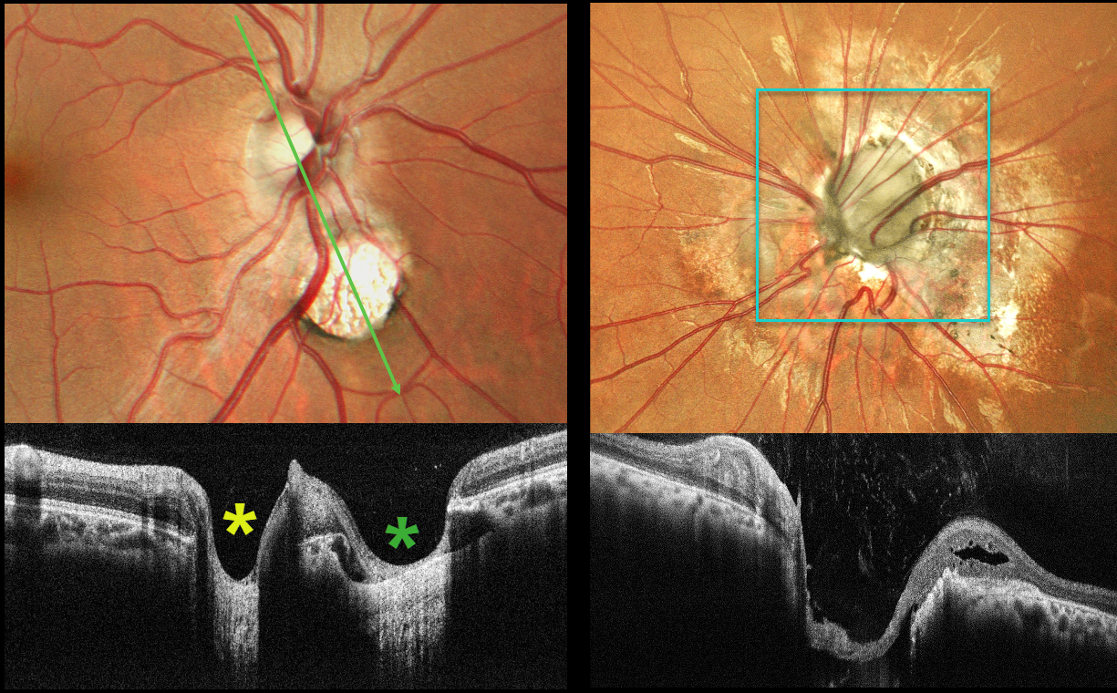
This 26YO male presented with a history of stable poor vision and inward deviation of his left eye since childhood. Vision was 20/20 OD and counting fingers in his esotropic, microphthalmic OS.
Color photography of the right posterior pole shows a round coloboma inferonasal to the nerve, giving the appearance of a pseudo-duplicated disc. OCT scanning through the nerve and coloboma reinforces the illusion of a second nerve head. The anomalous left nerve is enlarged and excavated, with numerous vessels radiating outwards from its inferonasal aspect.
Morning glory syndrome is typically unilateral and shares some similarities with optic disc coloboma and juxtapapillary staphyloma. This abnormality is named for its striking resemblance to the Morning Glory flower. Visual prognosis is usually poor. The condition can be associated with serous macular detachment. Neuroimaging should be considered to rule out transsphenoidal encephalocele and intracranial carotid artery dysgenesis, and fortunately, our patient’s imaging was negative. For a great review on the pathogenesis and treatment of maculopathy associated with cavitary optic disc anomalies, see Jain and Johnson, AJO 2014;158:423-435.
The coexistence of morning glory disc anomaly with microphthalmos, and contralateral chorioretinal coloboma in this case, may represent a continuum of optic fissure developmental defects with asymmetric expression, leading to a spectrum of cavitary optic disc anomalies in the same individual.
Pseudoduplication of the optic nerve occurs when a round choroidal coloboma or chorioretinal scar about the size of the optic nerve is associated with overlying radiating blood vessels (Bloom et al, Retinal Cases 2022;16:174-176). In our experience, this fake-out is most common from toxoplasmosis scars located near the optic nerve.



