

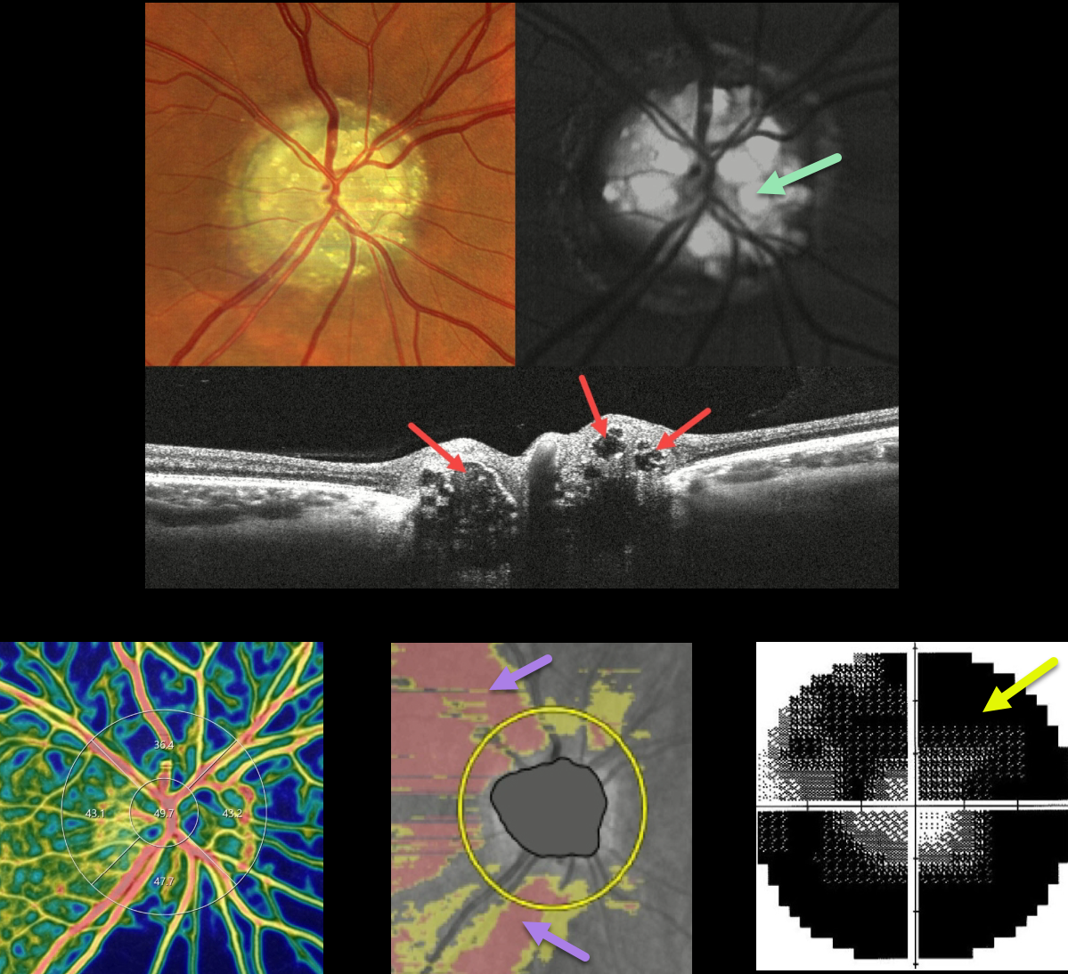
This 47YO female was examined for a routine eye examination. Although she initially denied any visual complaints, further questioning revealed a long history of peripheral vision loss. Vision was 20/20 in each eye, and the following findings were symmetrical and bilateral.
Color imaging of the right nerve shows severe optic disc drusen (ODD), which are hyper-autofluorescent. On OCT B-scan, the drusen appeared as relatively hyporeflective ovoid lesions within the substance of the nerve.
The drusen can be appreciated as blue flow voids on the OCT angiography density map. Diffuse retinal nerve fiber layer thinning is seen, and there is a severely constricted and depressed visual field. Her father was examined the same day and was discovered to also have ODD.
Learning Points:
ODD are refractile calcific concretions that form anterior to the lamina cribrosa. They are thought to be extruded, extracellular, calcified mitochondria and may be inherited as an autosomal dominant trait. Most patients are asymptomatic, but some can develop progressive visual field defects or central vision loss.
There is no treatment for the drusen themselves, although symptomatic macular neovascularization can be treated with thermal laser, photodynamic therapy, or anti-VEGF injections.



