

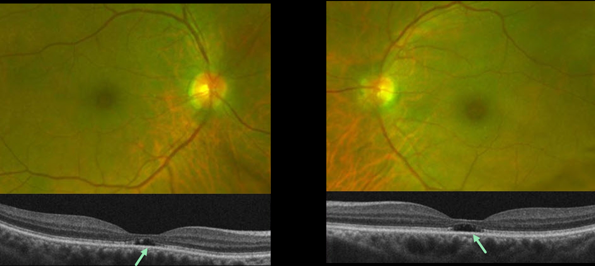
This 65YO presented complaining of difficulty seeing at a distance for some time. She gave a history of radial keratotomy in both eyes about 20 years prior. There was no significant past medical history or family history of eye disease. Vision was 20/60 OD and 20/70 OS.
Optos color imaging shows mild symmetrical pericentral pigmentary change. Fundus autofluorescence and fluorescein angiography were unremarkable (not shown). However, OCT B-scans show bilateral subfoveal loss of the outer retinal tissue external to the external limiting membrane.
An ERG showed rod receptor dysfunction and abnormal dark adaptation with delayed adaptation, while a multifocal ERG was consistent with normal cone function.
Our patient underwent genetic testing and was found to have a heterozygous mutation for FLVCR1. This gene is associated with autosomal recessive posterior column ataxia with retinitis pigmentosa.
A recent report by Kuehlewein et al (Graefe’s Arch Clin Exp Ophthalmol 2019;257:629-638) showed a variety of phenotypes, none of which matched our patient. It is therefore difficult to say whether our patient’s findings are isolated (given the lack of funduscopic, autofluorescence, and angiographic findings beyond the OCT outer defects) or represent an atypical manifestation (given the abnormal electrophysiology) of FLVCR1 disease.
She is scheduled to return for follow-up imaging and visual field testing.
Learning Points:
Outer foveal microdefects (Cohen et al, Ophthalmology Retina 2021;5:553-561) have been described in numerous conditions, including macular telangiectasia, tamoxifen use, ABCA4 disorders (cone-rod dystrophies, Stargardt disease, and fundus flavimaculatus), phototoxicity, trauma, and vitreomacular traction disorders.
In our experience, in patients with inherited retinal diseases (IRD), these findings are most common with a pathogenic ABCA4 mutation (Leng et al, Retina 2012;32:1411-1419 and Wang et al Invest Ophthalmol Vis Sci 2022;53(5):28). These defects, caused by focal loss of the outer retinal layers, leave an optically empty space which can be confused with subretinal fluid but are more of an optical gap within the outer retina.



