

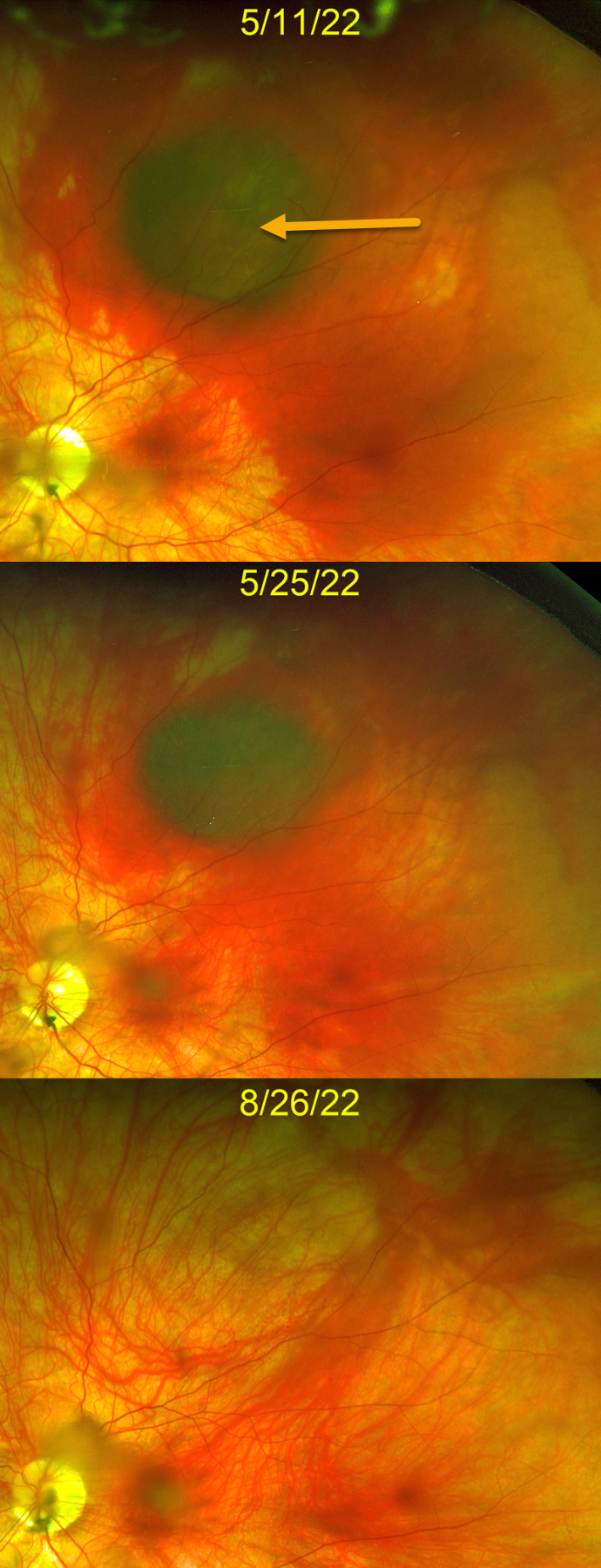
This 38YO healthy female presented with one day of peripheral vision loss in her left eye after an episode of forceful vomiting from a migraine headache. Vision was 20/30 OS.
Optos color imaging shows a dark, round, localized suprachoroidal hemorrhage in the superior midperiphery surrounded by subretinal blood. The right fundus was normal (not shown).
The blood gradually improved with observation, completely resolving over the next 3-4 months with return of 20/20 vision.
Learning Points:
Valsalva-induced retinal findings almost always present with sub-internal-limiting-membrane blood. Suprachoroidal hemorrhage most commonly develops intra- or postoperatively, following trauma or with thrombolytic agents (Ghu and Green, Survey Ophthalmology 1999;43:471-486).
Valsalva-induced choroidal hemorrhage is exceedingly rare, with only a handful of cases reported in the literature. As with other localized suprachoroidal hemorrhages, these lesions may simulate uveal melanoma (Marous et al, Retina 2018;12:336-341).



