

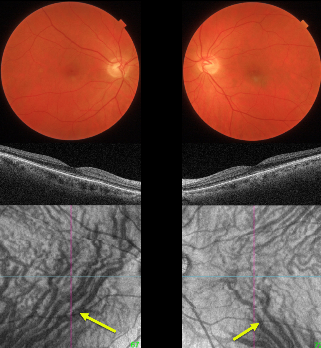
This 51YO minus 5 diopter myope presented without visual complaint for a routine yearly examination. Vision was 20/25 OU.
Both maculas are normal on fundus photo and B-scan OCT. However, en face OCT shows bilateral dilation of the inferior vortex system. The right inferior vortex also drains nearly the entire macula.
Learning Points:
The choroidal venous system, as with the neurosensory retinal vasculature, usually respects the horizontal meridian. With pachychoroid disorders, these anastomoses develop, and in this case, each inferior vortex system drains nearly the entire macula.
For a wonderful review highlighting the concept of venous overload choroidopathy, see Spaide et al, Progress Retinal Eye Research 2022;86:100973.
Pachychoroid, meaning “thick” choroid, is secondary to large dilated Haller vessels. Increased oncotic pressure from these dilated vessels can cause RPE dysfunction, serous detachments, and macular neovascularization.
Our patient has asymptomatic pachychoroid despite having a ‘normal’ choroidal thickness on B-scan OCT. The choroidal thickness depends on the refractive error. A high myope’s choroid should be quite thin, but if it appears as a ‘normal’ thickness, it is thick for that eye.
We also often see pachychoroid disease in elderly patients whose choroid is abnormally thickened for their age but appears ‘normally’ thick when not accounting for this. Although our patient’s retinal findings are completely normal clinically, these subclinical pachychoroid findings put her at risk for the various pachychoroid disorders
For an absolutely fascinating editorial discussing the ambiguity of how best to define what ‘pachychoroid’ actually means, see Spaide Retina 2021;41:231-237.



