

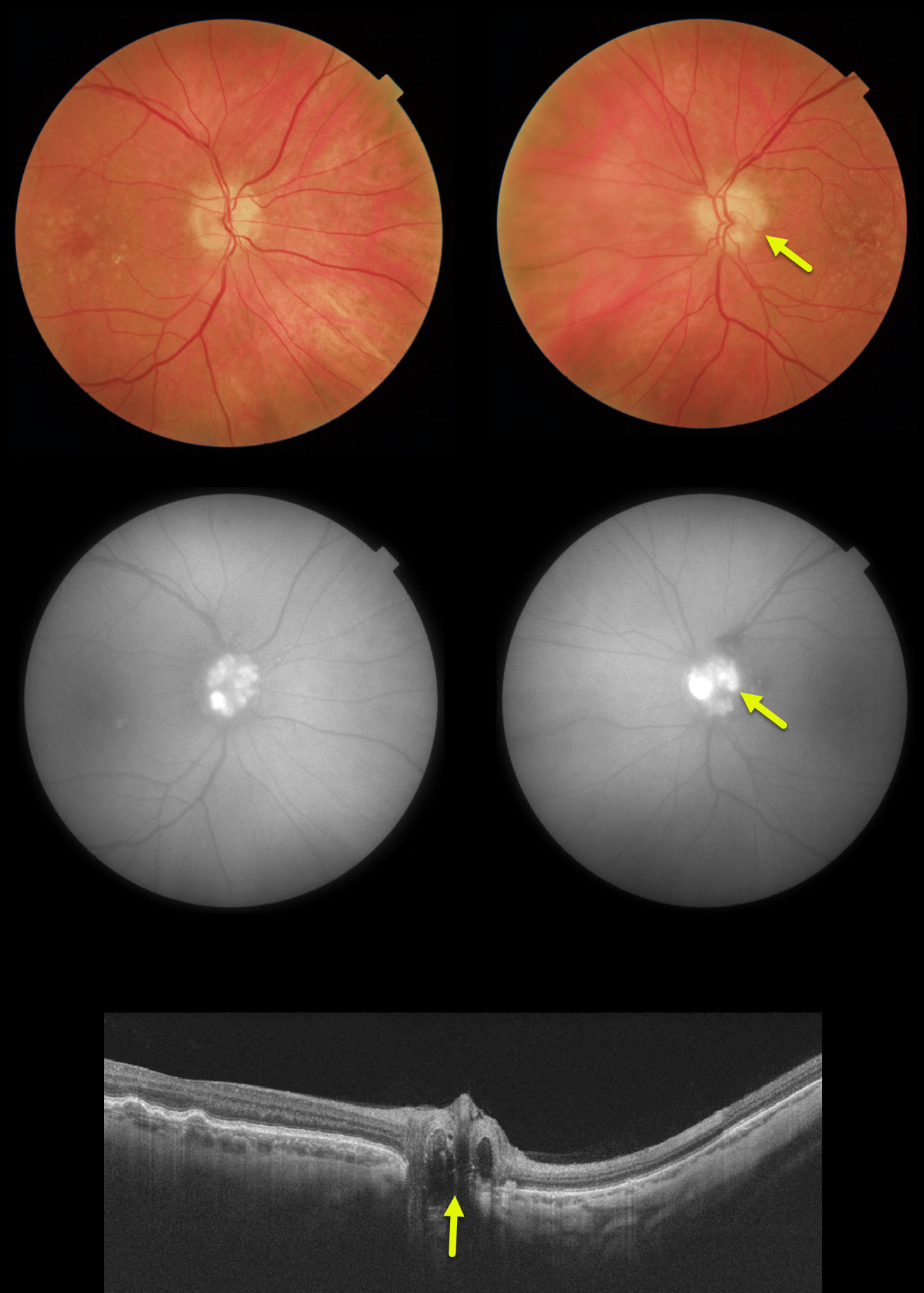
Our patient’s optic disc drusen were obvious clinically, on FAF and on OCT. In younger patients they are usually much more difficult to see and can be confused with papilledema.
We have found ultrasonography in younger patients cases to be totally unhelpful since drusen don’t calcify until later in life.
If fundus autofluorescence (FAF) is negative, the best clinical test in these cases is fluorescein angiography, which will show optic nerve leakage with papilledema. Eyes with drusen will show either no nerve leakage or nodular staining.
Learning Points:
Optic disc drusen are small hyaline bodies within the optic nerve that progressively calcify over time. They can be buried or superficial giving the appearance of pseudopapilledema. The nerve usually has a “lumpy-bumpy” appearance with normal overlying vessels. Retinal nerve fiber layer will usually show variably thinning.
FAF can show drusen hyperautofluoresence. Swept source OCT can occasionally visualize buried drusen.which can be either hypo- or hyperreflective.



