

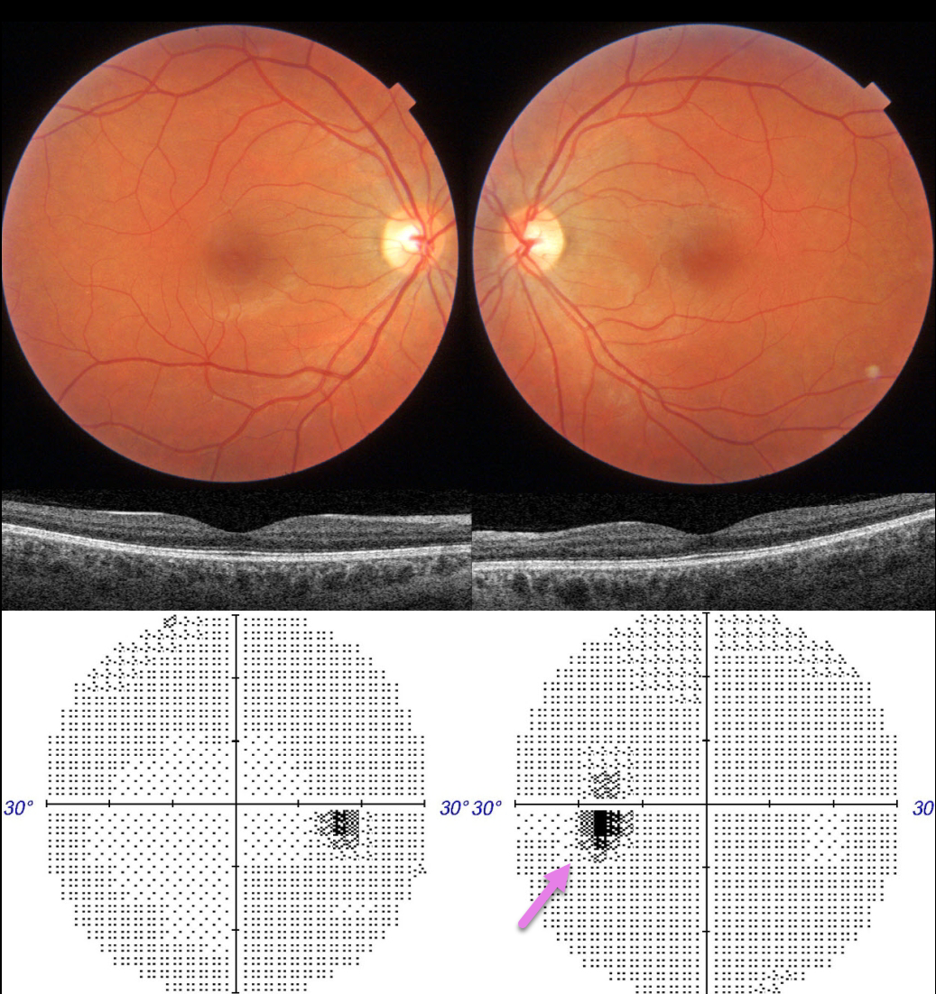
This 36YO female presented with 1 month of a paracentral scotoma in her left eye. There was no prior ocular or past medical history. Vision was 20/20 OU.
Ocular examinations were completely normal OU. Color imaging and OCT were normal. Visual fields were normal OD with a slightly enlarged blind spot OS.
Learning Points:
Multiple evanescent white dot syndrome (MEWDS) presents with usually unilateral idiopathic inflammation of the outer retina that typically occurs in young, healthy myopic females.
The granular fovea (Jampol spots, named after Dr. Lee Jampol, who first described this syndrome in 1984) is a classic pathognomonic finding that was not seen in this patient.
Acute cases will also have an enlarged blind spot, often with temporal visual field loss, due to peripapillary inflammation. The white dots disappear within a week or two, which is why the lesions are called “evanescent”. However, the blind spot may persist for many months, well after the fundus appearance has normalized.
This stage of MEWDS was originally called the Acute idiopathic blind spot enlargement (AIBSE) syndrome before Gass recognized that the two disorders were the same disease but with distinct findings at different points in time.
Observation for our patient was recommended. We expect her symptomatic enlarged blind spot will gradually resolve over the coming months.



