

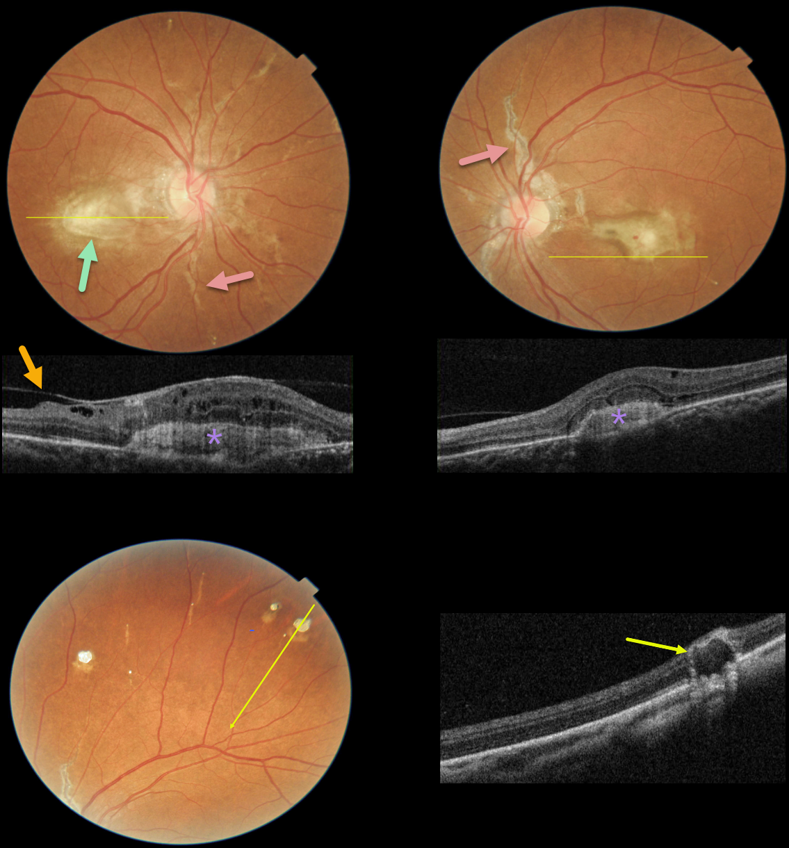
This 49YO female presented with one month of blurred vision. There was no past medical history except for a seizure disorder. Vision was 20/80 OD and 20/120 OS.
Fundus photographs show bilateral angioid streaks radiating from each optic nerve. Bilateral fibrosed subfoveal macular neovascularization (MNV) is present, and a small dot of blood is seen overlying the left macular lesion.
OCT scanning through the central right macula shows a hyperreflective, laminated MNV with overlying cystic retinal edema and a partially detached hyaloid vs macular pucker. OCT scanning through the inferior left macula also shows a hyperreflective MNV below the RPE associated with some overlying retinal thickening.
Numerous small subretinal comet-shaped lesions (‘comet rain’) are noted bilaterally. OCT scanning through one of these lesions shows a full-thickness hyporeflective lesion with a surrounding hyperreflective rim.
Learning Points:
Angioid streaks are orange-yellow, irregular, crack-like dehiscences in Bruch’s membrane associated with atrophic degeneration of the overlying RPE. They are often missed clinically since their color and orientation often blend in with the overlying retinal vessels.
Our patients’ lesions are easier to diagnose due to significant overlying RPE changes. The extent and prominence of the angioid streaks in our patient are pathognomonic for PXE, but less extensive streaks are seen in sickle cell disease and age-related macular degeneration.
These patients are at significant risk for macular neovascularization (MNV), which can be successfully treated with intravitreal anti-VEGF therapy.
The comet lesions are pathognomonic for PXE. These lesions develop from the outer retina as a neurodegenerative process and strongly resemble the hyporeflective core with a surrounding hyperreflective rim found in outer retinal tubulation (see Murro et al, Graefe’s 2018;256:1605-1614). Full-thickness lesions, as in our patient, were reported by Barteselli and Viola (Retina 2015;35:1051-1052).
Although our patient has no known personal or family history of PXE, her retinal findings indicate she has this disease. Her MNV appears chronic and fibrosed, and it is unclear why her symptoms were only present for one month. However, due to her recent symptoms and presence of overlying retinal fluid and blood, anti-VEGF therapy will be started.



