

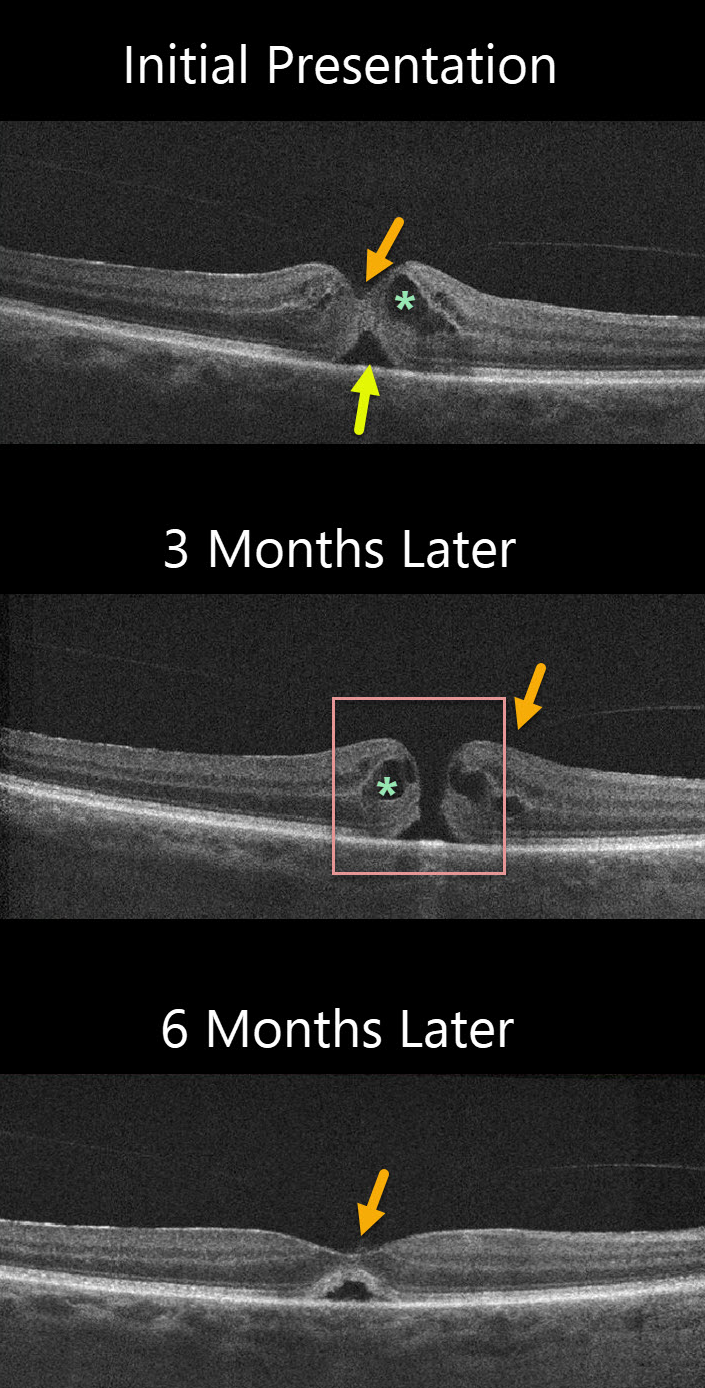
The patient was initially diagnosed with idiopathic cystoid macular edema (CME) and a small foveal detachment with vitreomacular adhesion (VMA). Vision was 20/50. Although the vitreous was attached centrally, a foveal depression remained which would be unusual if vitreomacular traction (VMT) were the cause.
The patient was observed without treatment, and 3 months later vision decreased to 20/60. There was a small full-thickness macular hole with an attached hyaloid. CME was still present. The patient elected to observe the hole.
Another three months later vision improved to 20/40 and the hole spontaneously closed with a normal foveal contour and resolved CME.
The central vitreous remained attached.
Learning Points:
The differential diagnosis for a localized serous foveal detachment include central serous chorioretinopathy, VMT, prior macular hole surgery, and resolved acquired vitelliform lesions.
This patient has a faintly visualized posterior hyaloid that is still attached centrally making VMT unlikely. The choroid probably has a normal thickness which makes central serous less likely, and moreover, the fellow eye is normal.



