

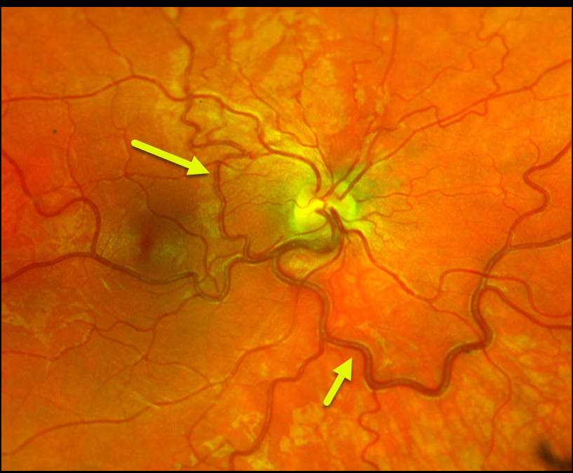
This healthy 24YO female presented for a general eye examination. There was no past ocular or medical history.
Color imaging of her right eye shows a large dilated retinal vein that drains the entire fundus except superonasally. It also crosses the temporal horizontal raphe into the superotemporal quadrant. Her left eye was normal.
Learning Points:
Retinal vessels virtually always respect the horizontal meridian. Retinal venous malformations (Pichi et al JAMA Ophthalmol 2018;136:372-379) are congenital anomalous vessels that by definition cross the horizontal.
Originally described by Gary Brown et al as congenital retinal macrovessels (Arch Ophthalmology 1982;100:1430-1436), these vessels are usually located in the macular region and are virtually always venous.
The findings in our patient are unusual, as the anomalous vessel supplies the majority of the retinal venous drainage.
These vessels are technically arterio-venous malformations (AVM) and are associated with venous anomalies in the brain (24% compared to up to 6% of the population). Brain MRI is therefore recommended to rule out any intracranial abnormalities. Neurologic imaging in our patient was fortunately normal.



