

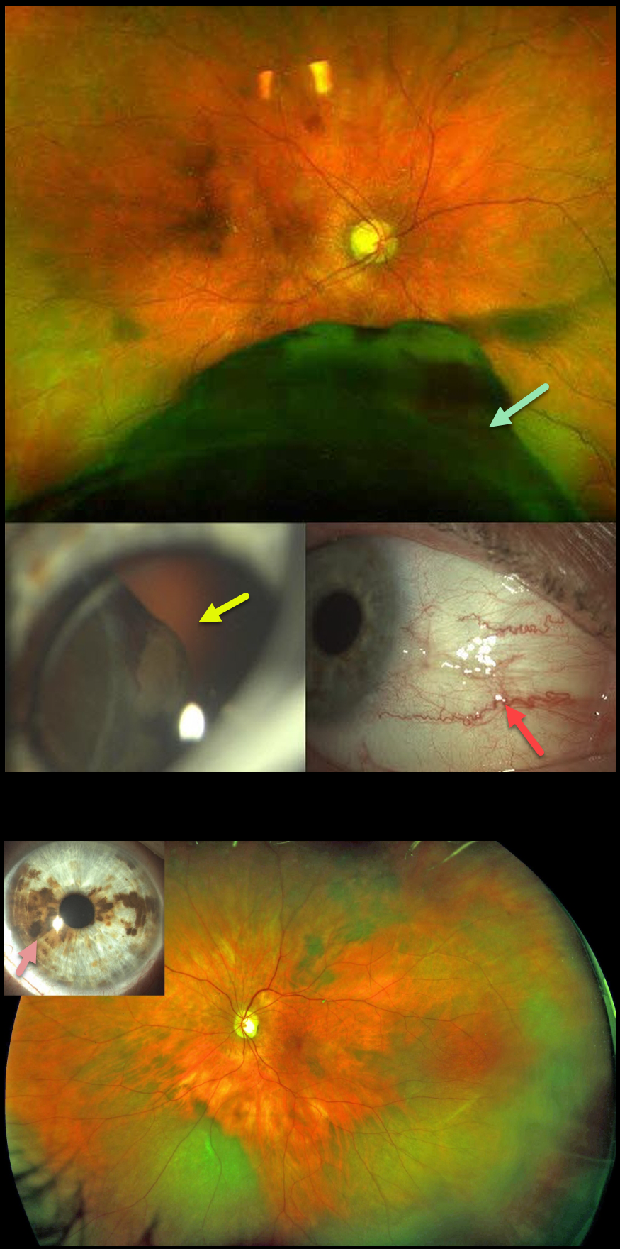
This 71YO female presented in January 2019 with one month of photopsias in her right eye. Optos imaging shows a large inferior malignant melanoma (MM) extending from the inferior retinal periphery into the posterior pole. The tumor was visible through the pupil at the slit lamp, and sentinel vessels were noted. Ocular melanocytosis was noted in the left eye.
She underwent primary enucleation of the right eye one month later. There was complete monosomy 3, and HLA-A*0201 testing was positive.
She subsequently developed a peripheral inferotemporal uveal malignant melanoma in her left eye (axial thickness 5mm, lateral dimensions 13.5 by 13 mm), which was treated with brachytherapy in March 2020. In October 2021, she developed liver metastases.
She was placed on systemic Nivolumab, in addition to external radiotherapy to the liver lesions. Vision is currently 20/20 OS with no signs of local recurrence.
Learning Points:
Our patient had a unique combination of unfortunate multiple risk factors for developing uveal malignant melanoma with metastatic disease (see Kaliki et al, Indian J Ophthalmol 2015;62:93-102).
Monosomy 3 is strongly associated with metastatic disease and melanoma-related mortality. The tumor suppressor gene BRCA1-associated protein 1 (BAP1) is mapped to chromosome 3p21.1, which is associated with autosomal dominant uveal MM and other primary cancers.
Choroidal melanocytosis is part of the oculodermal spectrum, which occasionally includes pigmentation of the globe or periocular skin (Nevus of Ota, melanosis oculi). There are increased dendritic melanocytes in the affected tissues following the distribution of the first and second branches of the trigeminal nerve. Patients need ongoing monitoring since about 1 in 400 will develop uveal melanoma. Those who develop melanoma are twice as likely to undergo metastasis when compared to patients with uveal melanoma and no pre-existing melanocytosis.
For a recent review of oculodermal melanocytosis, see Abdolrahimzadeh et al, Graefe’s 2023;261:291-301.



