

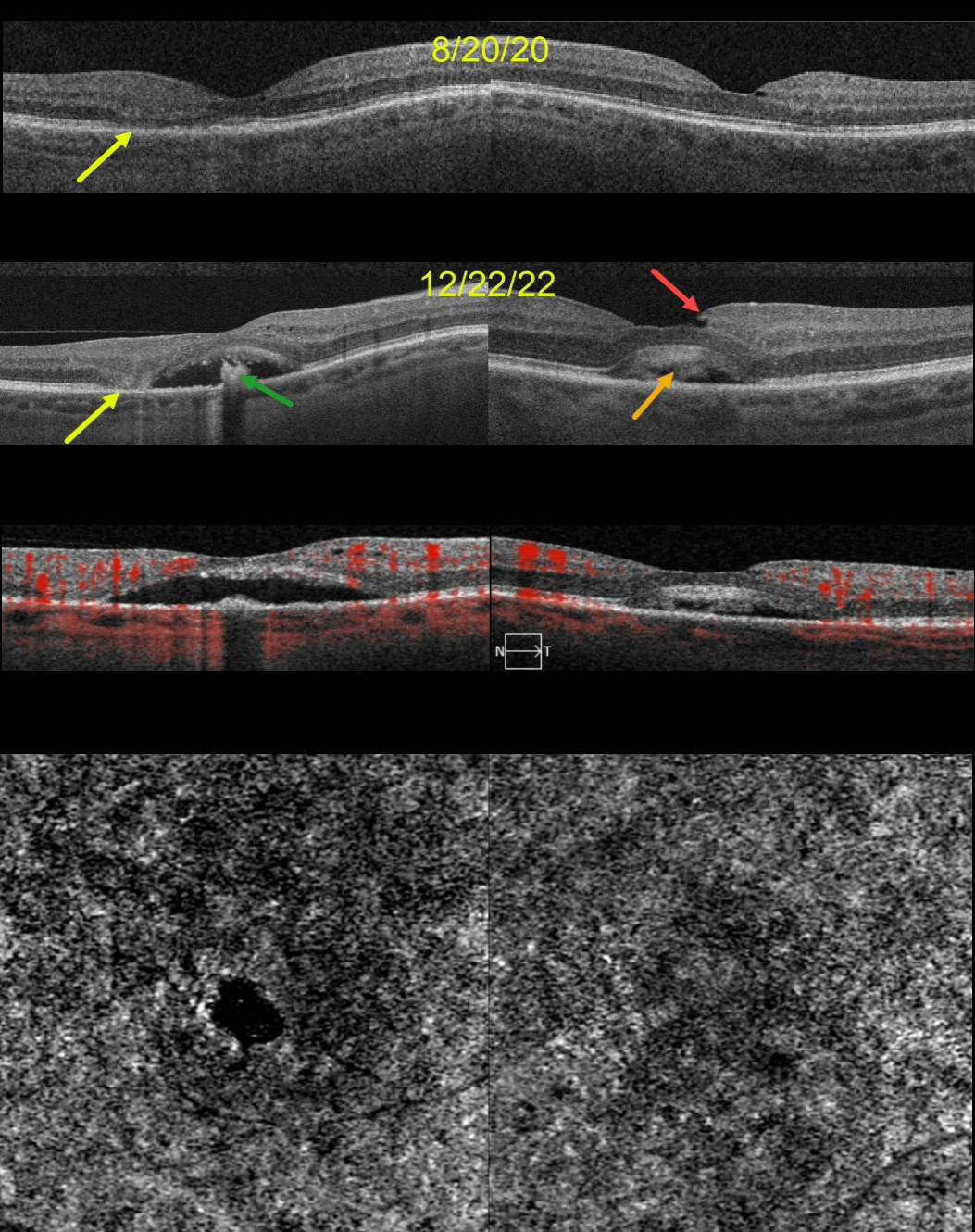
This 68YO female was seen on 8/20/20. Vision was 20/70 OD and 20/40 OS. Although there were no visual complaints, OCT scanning showed bilateral defects in the EZ bands, along with some temporal outer retinal atrophy OD.
She returned two years later, complaining of gradual vision loss OU, although vision was stable at 20/70 OD and 20/30 OS. OCT scanning shows bilateral acquired vitelliform lesions (AVL). The lesion OD is mostly scrambled with a hyporeflective space between the RPE and outer retina, along with a central hyperreflective clump on top of the RPE. There is now complete outer retinal atrophy temporally.
The AVL in her left macula shows variable hyperreflective material. An unrelated small partial lamellar macular hole is noted temporally. OCT angiography (OCTA) shows no flow signals from either AVL.
Learning Points:
Vision loss from AVLs most commonly results from lesions collapsing due to secondary atrophy or macular neovascularization, which can develop in about 8% of eyes (Balaratnasingam et al., AJO 2016;172:28-38).
The absence of OCTA flow signals in either eye confirmed that these lesions were avascular, and we continue to follow her.



