

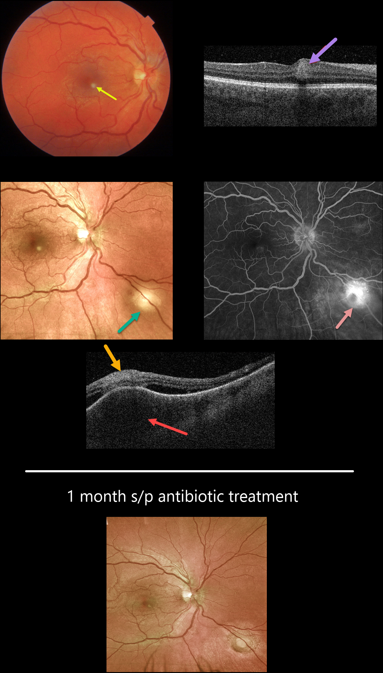
This healthy 27YO male presented with a few days of a paracentral scotoma in his right eye. Vision was 20/25 OD and 20/20 OS.
Color imaging shows a small inferonasal foveal area of inner retinal opacification. Swept-source OCT through this region shows inner retinal hyperreflectivity. An additional, larger subretinal lesion is noted in the inferonasal midperiphery. OCT scanning through this lesion shows marked choroidal thickening, overlying retinal disorganization and hyperreflectivity, and some surrounding subretinal fluid. Fluorescein angiography shows moderate leakage from this choroidal lesion.
On further questioning, the patient told us that he recently adopted 2 stray cats. He didn’t recall any cat scratches, rashes, or adenopathy, but Bartonella titers were markedly elevated (IgG 1:1280, IgM 1:400).
Following infectious disease consultation, he started a 6-week course of oral doxycycline and rifampin. Four weeks later, vision was 20/20, and both lesions had significantly improved.
Learning Points:
Bartonella Henselae is the bacterium associated with cat scratch disease. It is transmitted from cat fleas (carrying the bacterium) or from flea feces that can be present on the cat’s claws/teeth, and transferred to humans from a cat scratch or bite.
Common systemic findings include fever, malaise, and progressive lymphadenopathy. These findings usually occur within 1-2 weeks of a cat scratch/bite.
Ocular effects primarily include neuroretinitis with rare cases of Parinaud’s ocular glandular syndrome, or, as in our patient’s case, chorioretinitis.
In our practice, posterior segment Bartonella involvement most commonly presents with neuroretinitis. Our patient’s inflammation spared the nerve, with multifocal involvement including the inner retina and choroid.



