

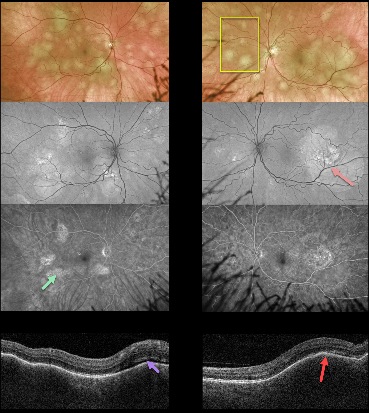
This 58YO female presented with 2 weeks of blurred vision in her left eye. There was no past medical history. Vision was 20/40 OD and 20/200 OS.
Optos color imaging shows multifocal, creamy-yellow subretinal lesions bilaterally. The lesions are more confluent, extending beneath the left fovea.
The smaller of these lesions is fairly silent on fundus autofluorescence, although the larger ones show hyper-FAF. These lesions angiographically show variable late staining with window defects.
Swept-source OCT shows bilateral thickened and undulating choroid with trace overlying subretinal fluid.
She was referred to the emergency room and was subsequently diagnosed with widely metastatic lung adenocarcinoma.
Learning Points:
Although uveal melanoma is the most common primary intraocular malignancy, metastatic choroidal tumors are the most common intraocular malignancies. Lung and breast cancer are the most common sources.
The extensive multifocal lesions in each of our patients’ eyes are unusual, since most patients have 1-2 choroidal lesions (Shields et al Retina 2020;40:204-213).



