

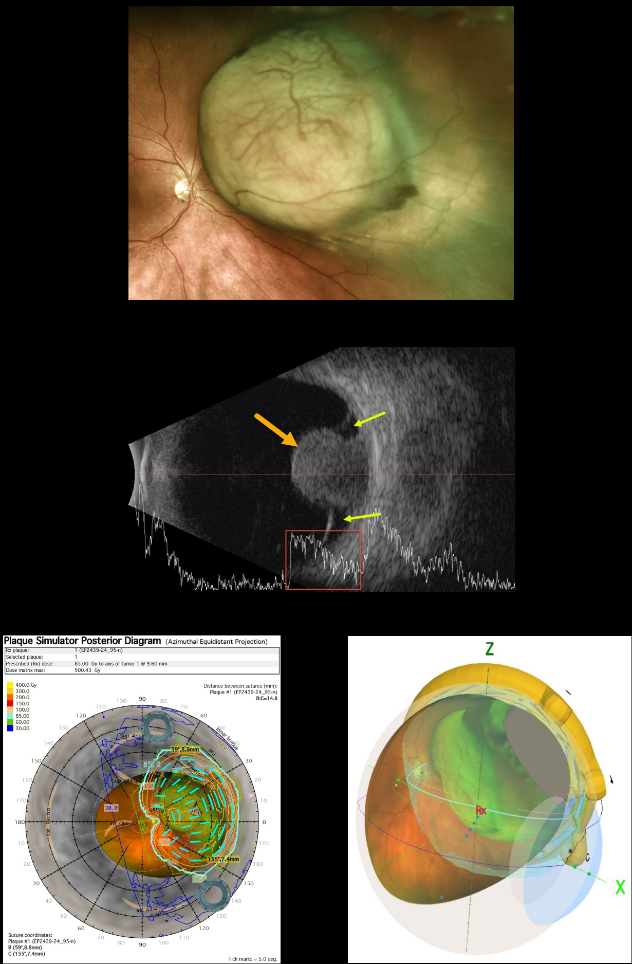
This 63YO female presented with several weeks of peripheral vision loss in her left eye. Vision was 20/40.
Optos color imaging shows a large, mostly amelanotic, mushroom-shaped choroidal lesion extending from just nasal to the nerve into the midperiphery. Intrinsic large vessels are noted. The lesion measured 11.5 × 14.5 mm in lateral dimensions.
Ultrasonography shows a mushroom-shaped choroidal tumor that measured 9.6mm in thickness. Localized subretinal fluid is seen on either edge. The lesion has moderate internal reflectivity.
Plaque radiotherapy of the uveal malignant melanoma (MM) was performed. The plaque simulator images created by the radiation oncologist during the design of the custom plaque with radioactive I-131 seeds are shown.



