

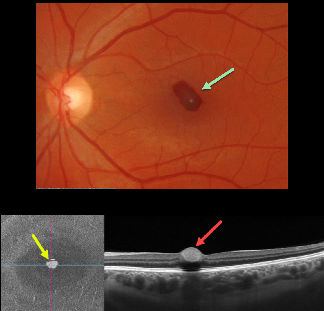
This 33YO female had a severe upper respiratory illness with intense coughing and sneezing. She woke one day earlier with a central scotoma in her left eye.
Optos color imaging shows a teardrop-shaped sub-internal limiting membrane (ILM) hemorrhage. Swept-source OCT B-scan shows a hyperreflective sub-ILM lesion with underlying shadowing. En face scanning of the inner retina provides a unique perspective on the blood.
Learning Points:
Valsalva hemorrhages are usually in the sub-ILM space. These hemorrhages virtually always resolve without sequelae, although the sub-ILM blood in severe cases can be drained into the inferior vitreous by creating a small ILM opening using either a thermal or Nd:YAG laser.



