

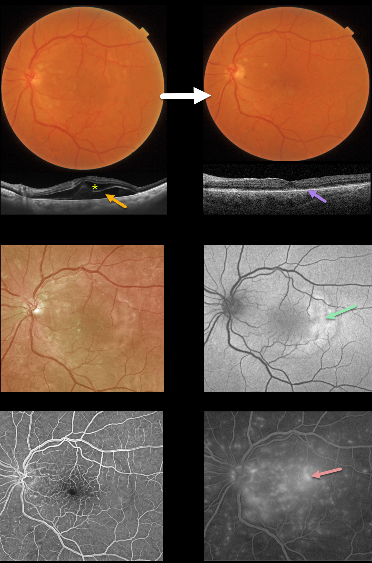
This 71YO male presented with decreased vision in his left eye one day following a motor vehicle accident with airbag deployment. Vision was 20/100.
Color photography shows variable faint patches of outer retinal whitening. Swept-source OCT shows outer retinal and subretinal fluid.
Optos fundus autofluorescence (FAF) shows temporal macular hyper-FAF. Fluorescein angiography shows early dots of subretinal hyperfluorescence with late deep leakage extending beyond the arcades.
Two weeks later, he was subjectively much improved, although he was bothered by metamorphopsia and some paracentral scotomas. Vision was 20/60. The macular pigmentary changes improved funduscopically. All macular fluid resolved, but the outer photoreceptor bands were disorganized and thinned.
Learning Points:
Posterior segment findings from blunt ocular trauma most commonly include commotio retinae or choroidal rupture.
Our patient’s unusual findings were from a contusive injury to the choroid and RPE. RPE dysfunction led to fluid accumulation in the subretinal space and the outer retina.
A strikingly similar case was reported by Mishra et al (Journal of Vitreoretinal Diseases 2021;5:165-169).



