

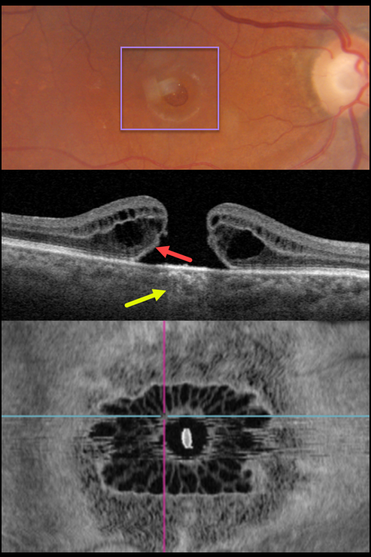
This 81YO female presented with 20/200 vision in her right eye from a large full-thickness macular hole.
Swept-source OCT shows the classic appearance of a hole from a foveal dehiscence with cystic thickening. Outer retinal bands are noted along the posterior aspect of the hole’s elevated edges, indicating the lack of true tissue loss. This retinal defect results in increased light transmission into the choroid. En face imaging shows a unique and beautiful perspective of the hole.
Vitrectomy surgery was recommended.
Learning Points:
As described by Govetto et al (AJO 2020;212:43-56), idiopathic macular holes have a sunflower appearance showing features of both exudative and tractional cystoid spaces. The macular hole is hyporeflective centrally with multiple hyporeflective cystoid spaces radiating from the center.



