

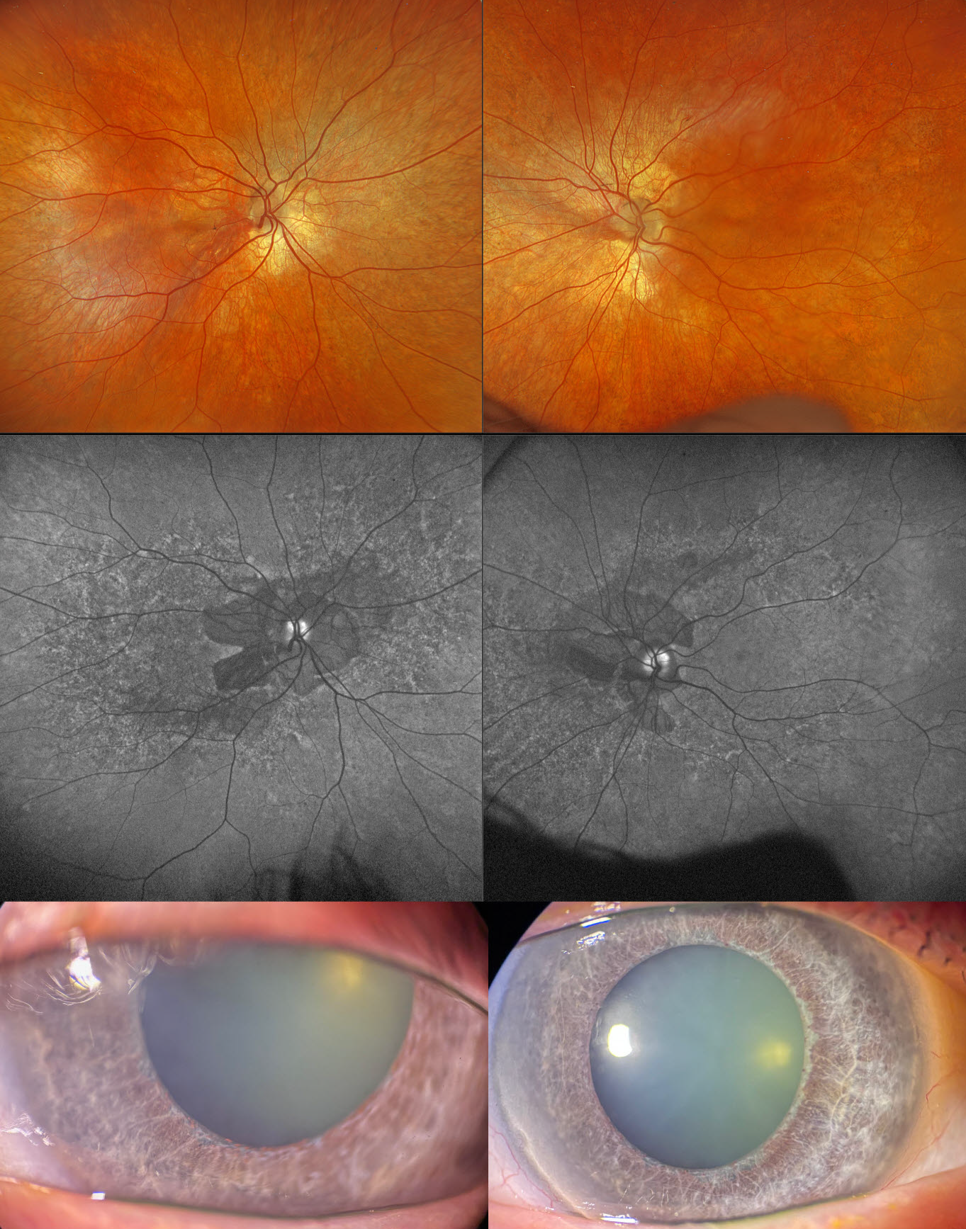
This 59YO male with type 2 diabetes was examined for his yearly screening. He has a known history of congenital rubella with deafness. Vision was 20/30 OD and 20/25 OS.
Optos color RGB imaging shows a diffuse salt-and-pepper appearance in each fundus, with peripapillary atrophy. On fundus autofluorescence (FAF), the peripapillary atrophy is hypo-FAF with a speckled hyper- and hypo-FAF appearance to each posterior pole. Slit lamp photographs show diffuse iris atrophy with a relative lack of pigmentation. Triton swept-source OCT shows variable outer retinal thinning with ellipsoid zone disruption.
Learning Points:
The classic ocular findings in congenital rubella include cataracts, pigmentary retinopathy (“salt-and-pepper” fundus), congenital glaucoma, and microphthalmia. Nuclear cataracts are the most common ocular abnormality, although our patient had just trace nuclear sclerosis. Pigmentary retinopathy is characterized by diffuse mottling of the retinal pigment epithelium, which is best visualized with FAF. Systemic associations include sensorineural hearing loss and congenital heart defects.



