

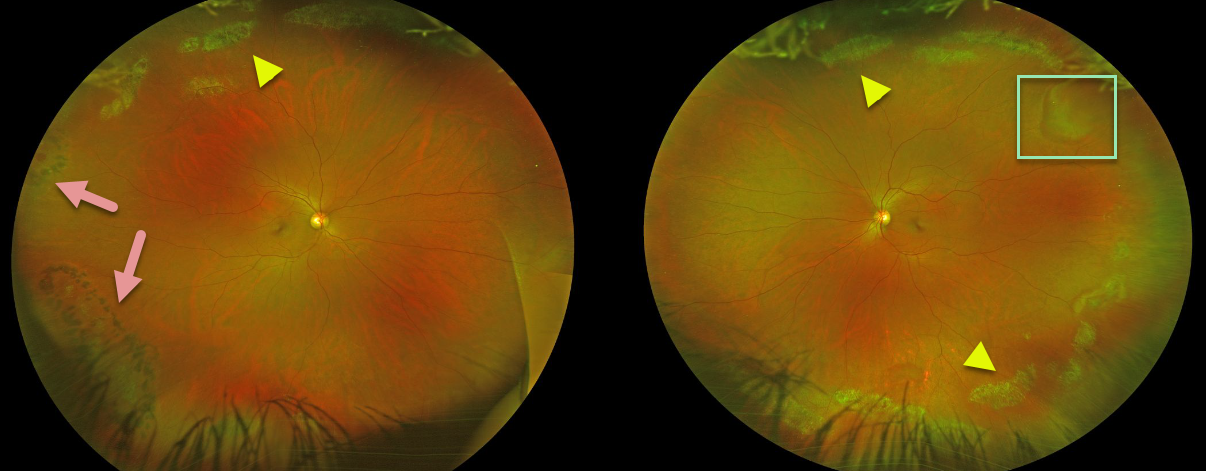
Beautiful Optos images of this patient demonstrate extensive bilateral lattice degeneration.
Prophylactic laser was placed at an outside institution around some of the lattice lesions in the right eye inferotemporally and around an atrophic hole temporally.
There is a pseudo-retinal tear superotemporally in the left eye from a patch of white without pressure.
Learning Points:
White, fibrosed vessels often criss-cross the lesions, which lead to the name, lattice. There is vitreous liquefaction overlying the thinned retina with a firm vitreo-retinal adhesion along the borders of the lesions. This firm vitreoretinal adhesion, along with the often present atrophic holes, is what can predispose these eyes to retinal detachment.
Retinal detachment occurs in 1 in 10,000 people per year. Lattice is found in 10% of the general population, especially in high myopes. Since about one-third of patients with retinal detachment have lattice as the cause, the question always arises, especially in patients like this: should prophylactic retinopexy be performed? The short answer is that asymptomatic lattice generally should not be treated.



