

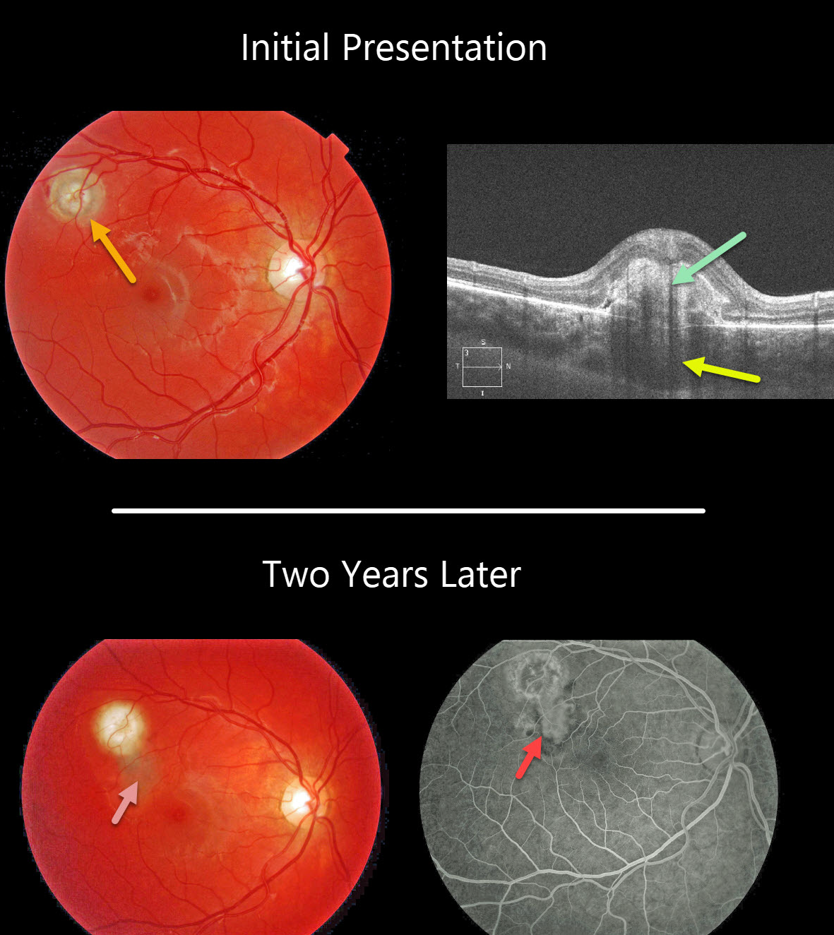
This patient’s fundus photo shows a focal yellow-white nodular lesion of the right eye. OCT shows the subretinal hyperreflective material and an underlying choroidal granuloma.
Two years later, the patient subsequently developed an active macular neovascularization along the inferior edge of the original lesion, seen better on fluorescein angiography.
Learning Points:
Choroidal granulomas usually appear as solitary creamy white, round subretinal lesions. They are hyporeflective on OCT imaging (Invernizzi et al, Retina 2015;35:525-531). Causes include sarcoidosis, tuberculosis and VKH. They can occasionally cause exudation that responds to anti-VEGF injections or photodynamic therapy.



