

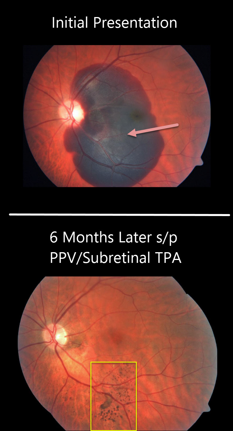
The large submacular hemorrhage is likely from a macular neovascularization from polypoidal choroidal vasculopathy.
The patient was treated with vitrectomy, subretinal tissue plasminogen activator (TPA), and fluid air exchange. The TPA helped liquefy the clot, which was then hydraulically displaced inferiorly outside the macula by the gas bubble.
At six months post-op, the central macula is remarkably normal with multifocal clumps of subretinal pigment more inferiorly.
Learning Points:
Multifocal subretinal pigment is a classic finding for regressed subretinal blood. This pigment is hypoautofluorescent without RPE atrophy (see Hussnain et al, Retina 2019;39;1925-1935).



