

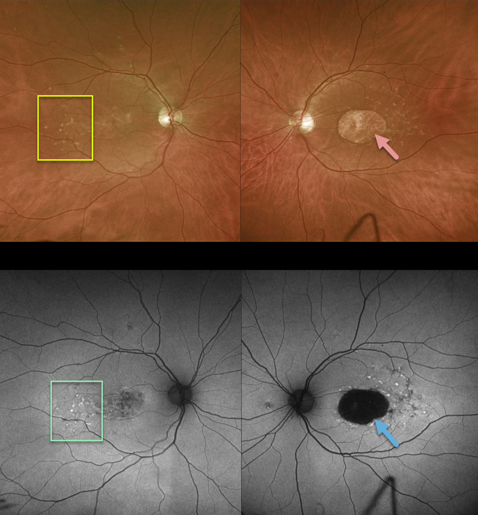
This patient has bilateral 20/400 vision due to classic findings from Stargardt disease, including yellow subretinal flecks and outer retinal/RPE atrophy.
FAF primarily displays hyperfluorescence from RPE lipofuscin. Since Stargardt is essentially a lipofuscin storage disease, the subretinal flecks are hyperautofluorescent and the areas of RPE loss are hypoautofluorescent.
Learning Points:
Stargardt disease is an autosomal recessive disorder caused by a mutation in the ABCA4 gene. The ABCA4 protein is located in the photoreceptor outer segments and is involved in the recycling of 11-cis-retinal. The mutation of this gene causes accumulation of lipofuscin in the RPE, which eventually leads to photoreceptor and RPE degeneration.
Fluorescein angiography (not pictured) characterically shows a ‘dark choroid’ from the lipofuscin-engorged RPE blocking visualization of the underlying choroid.
Stargardt is one of the macular diseases where the vision loss is often much worse than expected from the fundus appearance.



