

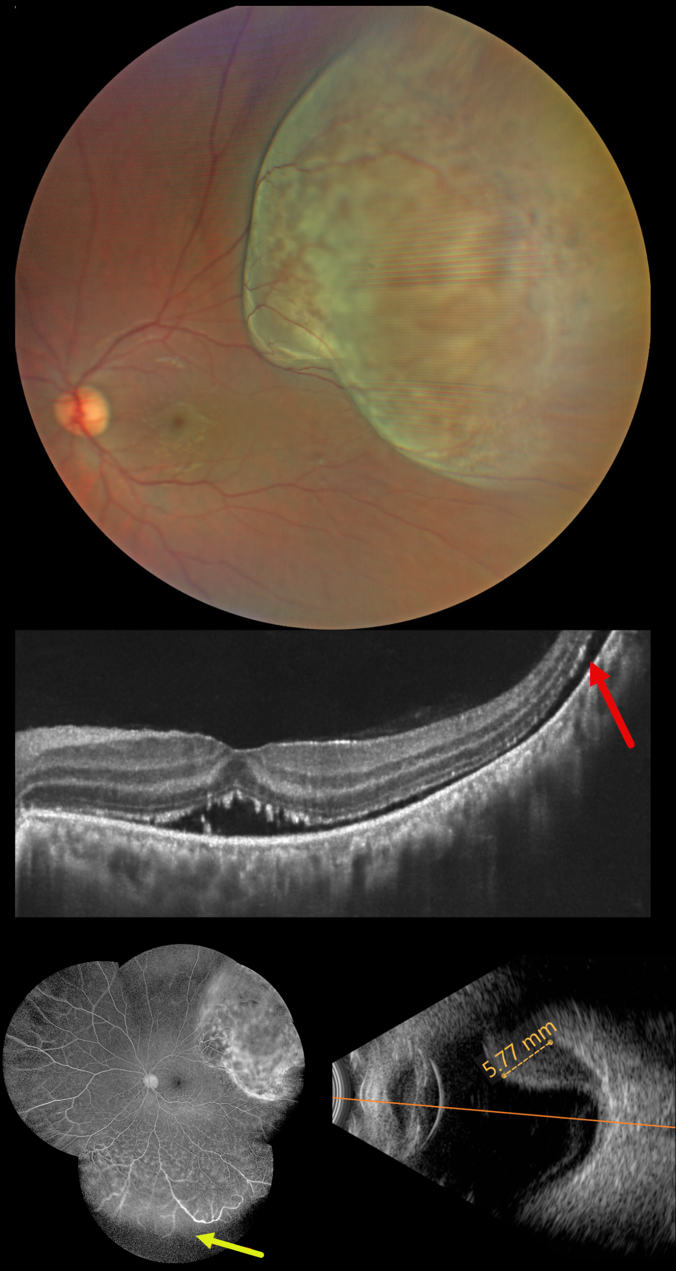
This healthy 36YO male presented with two weeks of vision loss in his left eye. Vision was 20/20 in his normal OD and 20/40 OS.
Fundus photography shows an elevated, pigmented, solid choroidal tumor, which spares the macula but occupies nearly the entire superotemporal quadrant. OCT scanning shows shallow subretinal fluid tracking from the tumor into the fovea with hyperreflective shaggy outer segments. Fluorescein angiography shows leaking intrinsic tumor vascularity with inferior peripheral retinal capillary loss. The tumor, which measures 5.77mm in axial thickness, shows moderate internal reflectivity on B-scan ultrasonography.
Learning Points:
Uveal melanoma is a malignant tumor arising from melanocytes in the uveal tract (iris, ciliary body, or choroid). The choroid is the most common site, accounting for 90% of uveal melanomas. Uveal melanoma is the most common primary intraocular cancer in adults, whereas metastatic disease is the most common intraocular malignancy. The liver is the most common metastatic site, although metastatic disease is rare on presentation. Metastases are more common in large tumors, with BAP1 mutations, and with certain tumor-related genetic markers (including positive PRAME and Class 2).
He was immediately referred to an ocular oncologist for further evaluation and treatment, including genetic testing, metastatic workup, and plaque brachytherapy. One month postoperatively, the tumor regressed to 4.6mm in thickness with decreased exudative fluid (not shown).



