

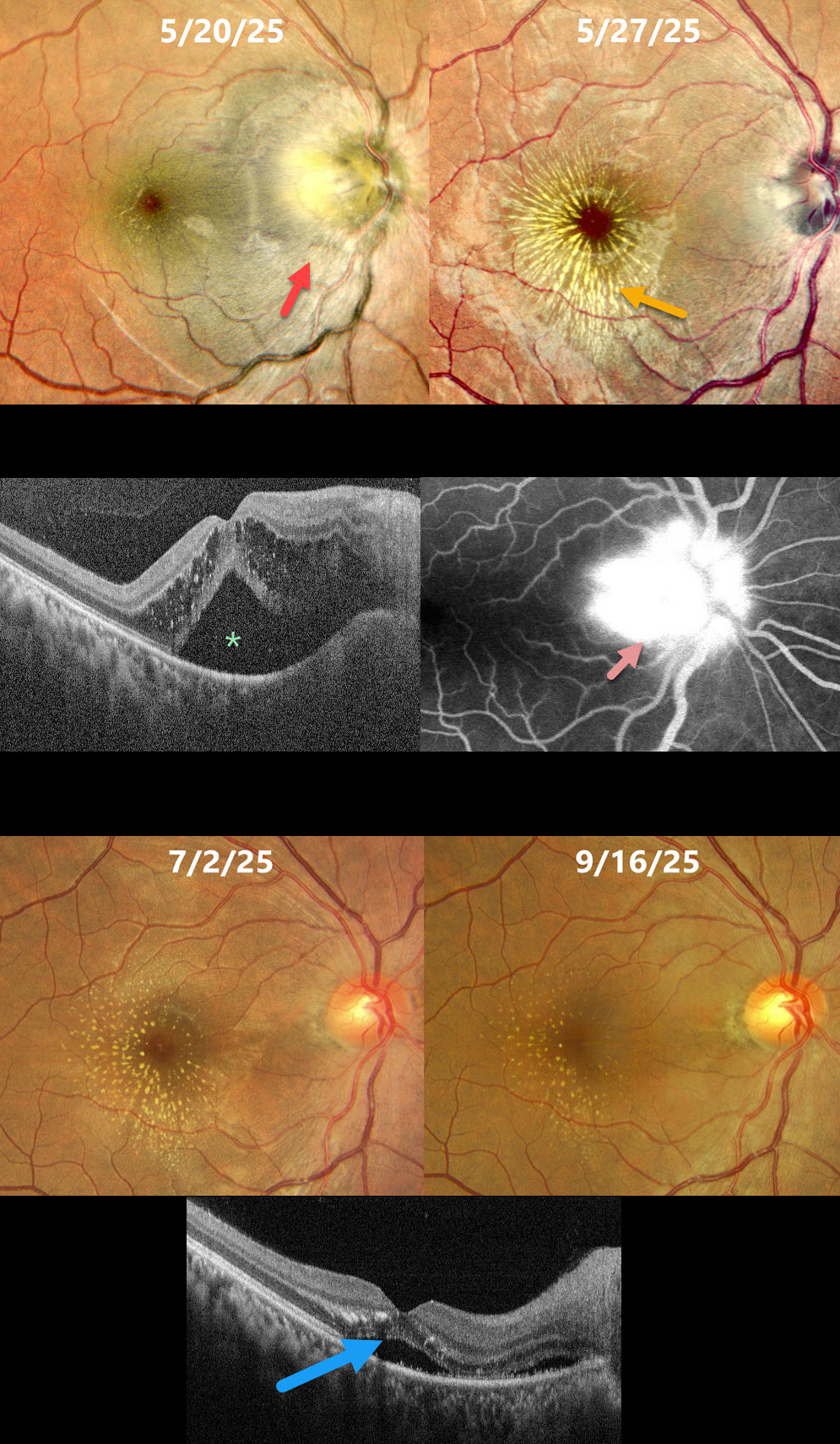
This healthy 32YO female presented with a 10-day history of headaches, followed by vomiting and blurred vision in her right eye. She also reported a rash on her hands and legs that appeared before her visual symptoms. Vision was 20/300 OD and 20/20 in her normal OS.
Pseudocolor SLO imaging shows a swollen optic nerve, a serous detachment extending through the fovea, and some mostly temporal radiating foveal lipid. OCT scanning shows outer nuclear layer fluid with hyperreflective lipid deposits and subretinal fluid. Fundus fluorescein angiography shows marked disc leakage. Infectious disease consultation revealed a negative infectious workup, and she was started on intravenous followed by a tapering dose of oral steroids.
One week later, vision was 20/300 with a striking new lipid star from lipid in the outer plexiform layer. There is a marked reduction in the intra- and subretinal fluid. Her findings continued to improve, and 4 months following presentation, vision was 20/20 with faint residual lipid.
Learning Points:
The diagnosis for a unilateral swollen nerve is extensive. However, it becomes much smaller when associated with acute inner retinal ischemia (not seen in our case) and later lipid exudation within Henle’s layer (neuroretinitis), including cat scratch (Bartonella) and syphilis. When no underlying cause is found, as in our patient, the entity is called Leber’s idiopathic stellate neuroretinitis.



