

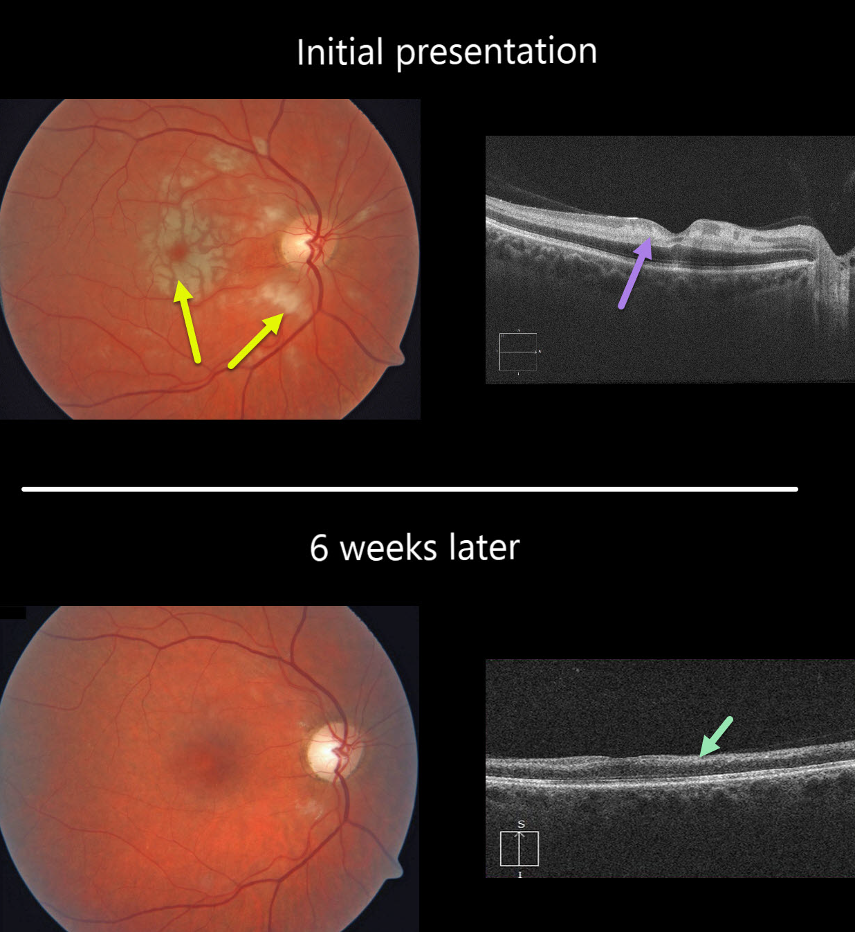
This healthy 49YO female presented with severe, sudden vision loss in her right eye.
There were multiple white macular patches similar to Purtscher’s retinopathy, but without retinal hemorrhages.
Optical coherence tomography (OCT) showed multiple mid-retinal hyperreflective paracentral acute macular maculopathy (PAMM) lesions. Since we felt this could represent an incomplete central retinal artery occlusion (CRAO), we sent her immediately to the nearest stroke center for further assessment.
At her six-week follow-up, the retinal appearance virtually normalized. However, OCT showed severe diffuse thinning of the inner non-photoreceptor retinal layers consistent with a resolved CRAO. Her stroke workup was unremarkable.
Learning Points:
Purtscher’s retinopathy was first described by Otmar Purtscher in 1910. Although originally described in a man who fell from a tree with cranial trauma, these white patches of retinal ischemia and hemorrhages are found in numerous other etiologies, including pancreatitis, chest trauma, and collagen vascular diseases.
The findings are called Purtscher’s retinopathy when due to trauma, and Purtscher ‘s-like retinopathy when seen from other causes.
The underlying cause of Purtscher’s is unknown, but is thought to involve leukoembolization due to complement activation. The areas of ischemia include more superficial nerve fiber layer infarcts (cotton wool spots) and deeper ischemia involving the middle retinal layers (PAMM).



