

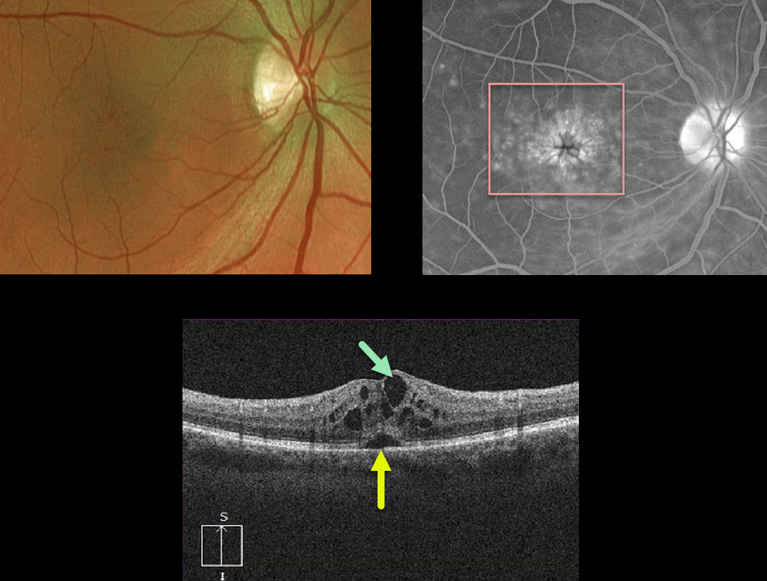
This is a classic case of cystoid macular edema (CME) following cataract surgery, otherwise known as the Irvine-Gass Syndrome.
The decompensated inner blood retinal barrier of the perifoveal capillaries allows fluid to leak into the surrounding outer plexiform (Henle’s) layer, causing the classic radiating petaloid pooling on fluorescein angiography.
OCT shows intraretinal edema with cystic spaces and some foveal subretinal fluid.
Learning Points:
Dr. Irvine was the first to describe the condition in 1953 (AJO 1953;36:599-619), and Dr. Gass expanded on this entity in his classic paper (Arch Ophthalmology 1966;76:646-661).
Clinically, at the slit lamp, the cystic changes are best visualized with a contact lens examination (this was how Drs. Irvine and Gass figured things out 60-70 years ago!), although this is rarely currently utilized due to the wide availability and superiority of OCT.
For a great explanation for differentiating exudative from tractional cystic spaces with multimodal imaging, see Govetto et al AJO 2020;212:43-56.



