

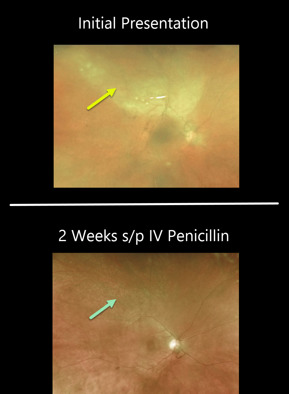
This 46YO female presented with worsening pain and vision loss in her left eye despite being treated two days earlier by the emergency room for conjunctivitis. In our office, vision was counting fingers OS with a granulomatous panuveitis.
There is a creamy-white area of confluent retinitis superiorly. The image is hazy due to vitritis.
Stat bloodwork revealed a positive RPR and FTA-ABS. She denied any other symptoms or known history of syphilis.
Patients also need to be evaluated for HIV since syphilis and HIV infections often coexist. Fortunately, our patient was HIV negative.
After 2 weeks of parenteral penicillin, the panuveitis had improved markedly, and the retinitis had completely resolved.
Learning Points:
Ocular manifestations of syphilis vary widely and include virtually all variations of anterior and posterior segment inflammation, including a recently described presentation as the multiple evanescent white dot syndrome phenotype (see Russel et al Int Ophthalmol 2020;40:627-738).
Hence, the reason it’s been dubbed the great imitator or masquerader. One of the more common presentations is placoid chorioretinitis.



