

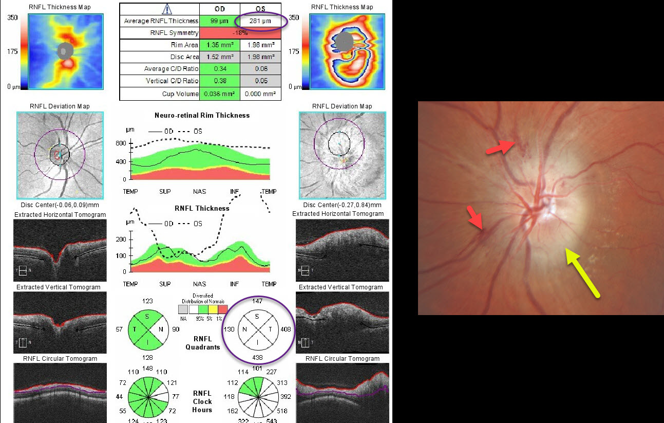
This patient presented with unilateral diffuse optic nerve edema with some splinter hemorrhages and possible inferotemporal lipid. Optic nerve OCT confirms markedly increased retinal nerve fiber layer thickness.
The differential diagnosis for a unilaterally acutely swollen nerve is quite long and includes arteritic (AION) and non-arteritic (NAION) anterior ischemic optic neuropathy, infectious causes (including syphilis and neuroretinitis), and inflammatory conditions (including sarcoidosis and optic neuritis). In this case, we diagnosed NAION.
Learning Points:
NAION presents with sudden painless unilateral central vision loss with an altitudinal defect. It is the most common acute optic neuropathy in patients over age 50.
It is thought to be caused by non-embolic occlusion of small branches of the short posterior ciliary arteries that supply the anterior optic nerve. It is associated with congenitally small and crowded nerves, optic disc drusen, medications (amiodarone and possibly phosphodiesterase inhibitors), hypertension, and diabetes. About 75% of patients have sleep apnea. Nocturnal hypotension or taking blood pressure pills at bedtime is also considered a risk factor.
Erythrocyte sedimentation rate and C-reactive protein should be ordered to rule out giant cell arteritis. There is no effective treatment.
Optic nerve edema resolves within a few months, often with secondary optic nerve pallor.



