

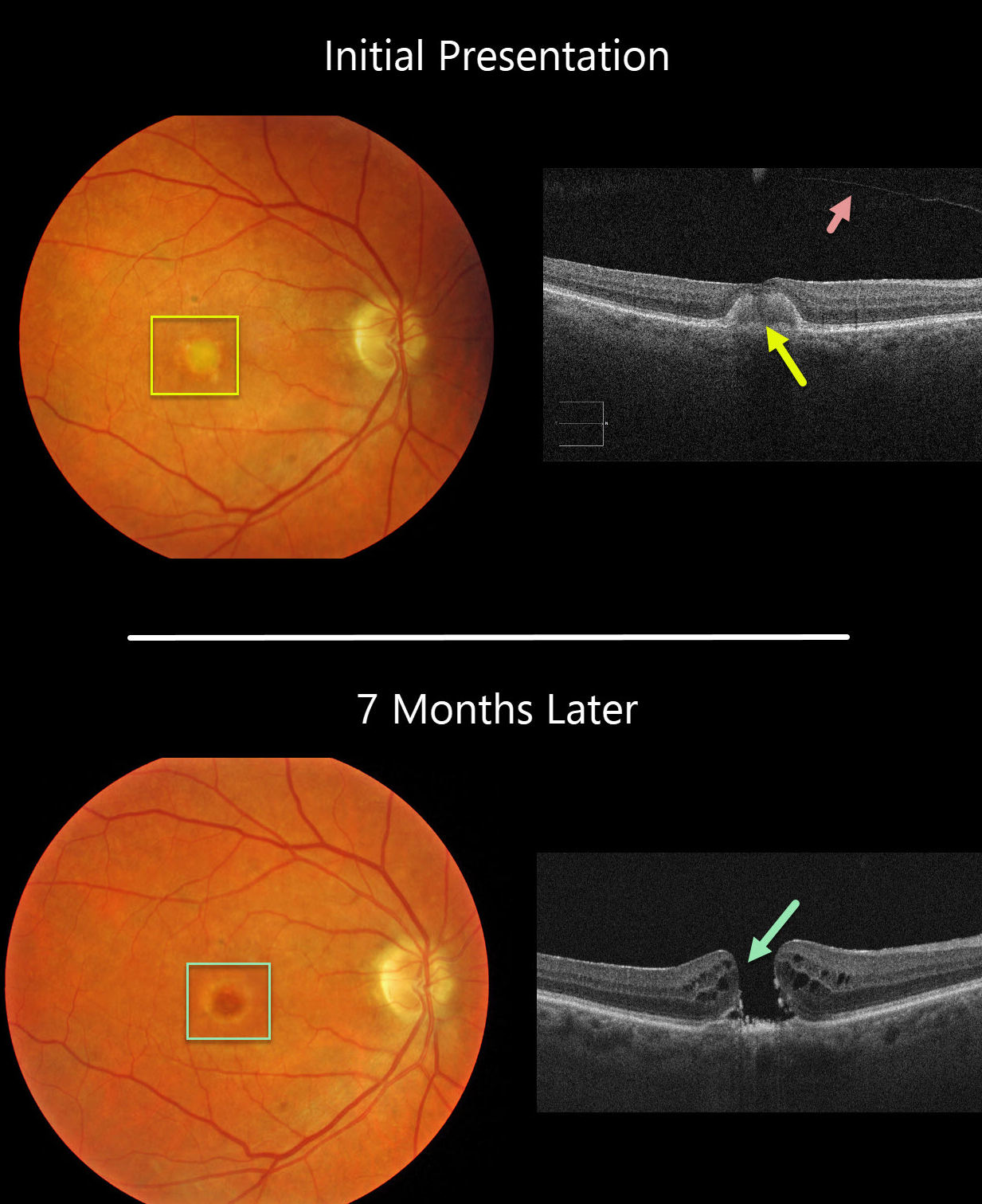
This 77YO female was initially examined with 20/60 vision OD, 20/30 vision OS, and bilateral vitelliform lesions (only right eye pictured) consistent with Adult Best (adult-onset foveomacular vitelliform dystrophy). A complete posterior vitreous detachment (PVD) was noted in her right eye.
Seven months later, vision decreased to counting fingers OD due to a new full-thickness macular hole. This patient was asymptomatic due to good vision in her left eye and has opted against surgery for now.
Learning Points:
Macular holes are most commonly caused by a partial PVD with active vitreomacular traction. However, they can occasionally develop without traction following a prior PVD or vitrectomy surgery. Macular holes have also been reported as a rare complication of adult Best’s disease.
Adult-onset foveomacular vitelliform dystrophy is associated with a mutation in the BEST1 gene, which encodes the bestrophin-1 protein.
Bestrophin-1, a calcium-activated chloride channel, is primarily found in the basolateral plasma membrane of the RPE. BEST1 mutations cause a variety of phenotypes, including autosomal recessive bestrophinopathy, best vitelliform macular dystrophy, and autosomal dominant vitreoretinochoroidopathy.



