

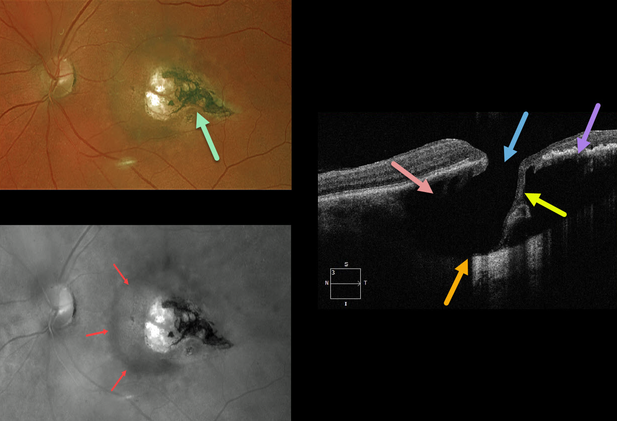
This 67YO female was referred with 20/30 vision and asymptomatic retinal findings. There was a tear-drop-shaped, variably pigmented chorioretinal scar in the temporal macula. The associated increased choroidal pigmentation is best seen in the red-channel image.
The OCT shows some unexpected findings. There is either a coloboma or posterior staphyloma causing marked posterior displacement of the sclero-choroidal junction. Temporally thinned retina dives posteriorly with an adjacent full-thickness retinal defect.
Temporally, the increased pigmentation of the scarring likely makes it difficult to see more posterior choroidal detail due to shadowing. Nasally, the faint, increased choroidal pigmentation also causes shadowing.
What do you think this is? Our best guess is a choroidal cavitation or an atypical colobomatous toxoplasmosis scar.
Learning Points:
For a great discussion on how to differentiate these infectious macular colobomatous lesions (congenital toxoplasmosis, Zika, and cytomegalovirus) from inherited dystrophies like North Carolina Macular Dystrophy (NCMD), see Kumar and Mahalingam AJO 2019;200:47-56. Also see the first OCT description of congenital toxoplasmosis macular lesions (Garg et al, Retina 2009;29;631-637).



