

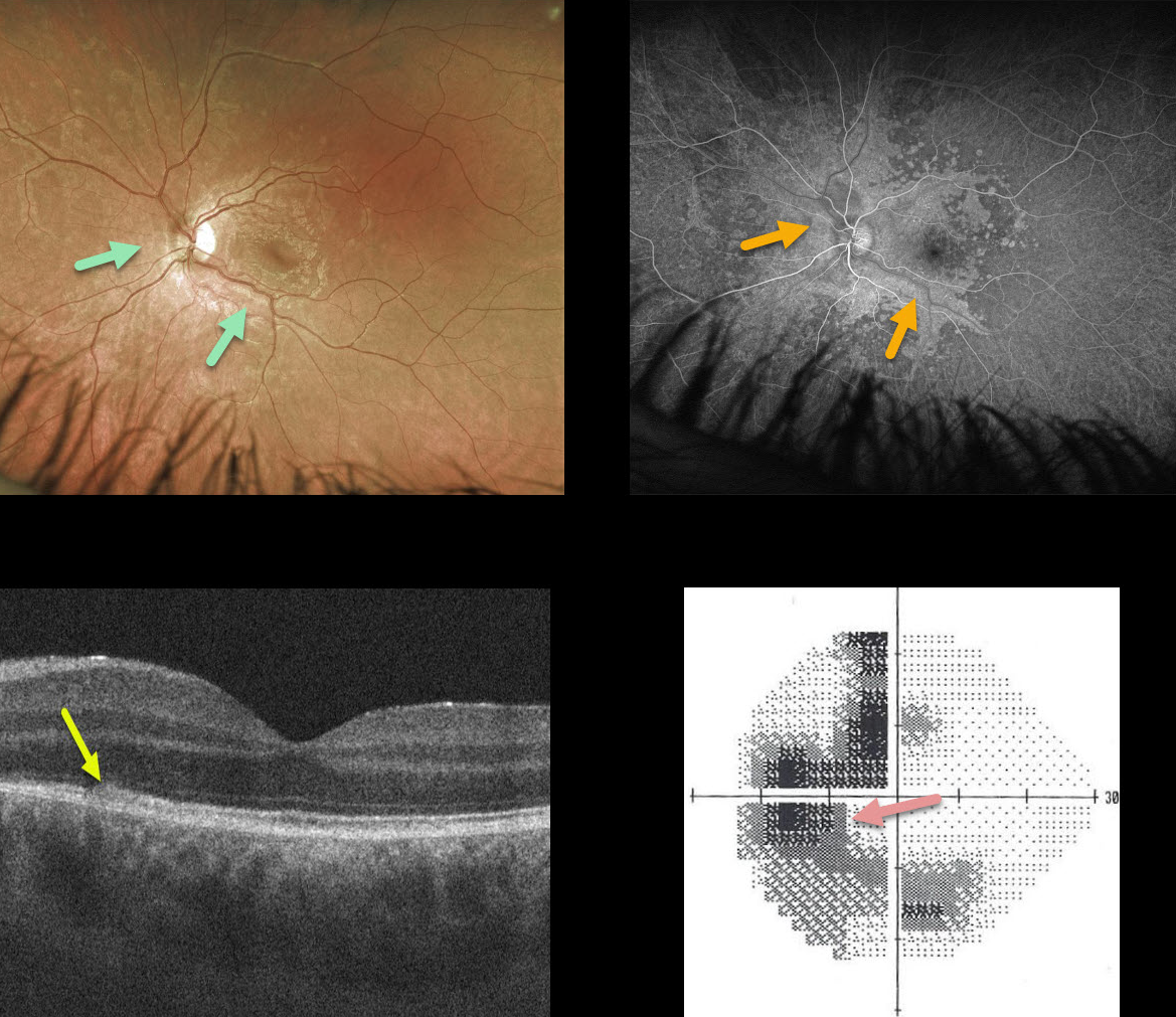
This 23YO patient presented with a few days of a temporal blurred spot in his left eye. Vision was 20/20 OD and 20/25 OS.
The left fundus has confluent outer retinal white spots centered around the optic nerve and extending into the macula. These lesions also had an unusual perivascular distribution more peripherally.
Fluorescein angiography shows hyperfluorescence staining in these lesions. Although this appearance suggested a possible vasculitis, angiography revealed absolutely no vascular changes. We are not sure why the outer retinal spots appeared in this vascular distribution.
OCT shows some hyperreflectivity and loss of detail of the ellipsoid zone and outer retina, and visual field testing shows an enlarged blind spot with temporal and inferior loss.
Learning Points:
We initially considered the possibility of syphilis given the pseudo-vasculitis and placoid confluence of the outer retinal lesions. However, the patient is in a long-term monogamous relationship and declined initial RPR/FTA testing.
Visual symptoms improved without any treatment. When the patient returned 6 weeks later, the white spots had disappeared clinically and on fundus autofluorescence, and his scotoma and symptoms had improved.



