

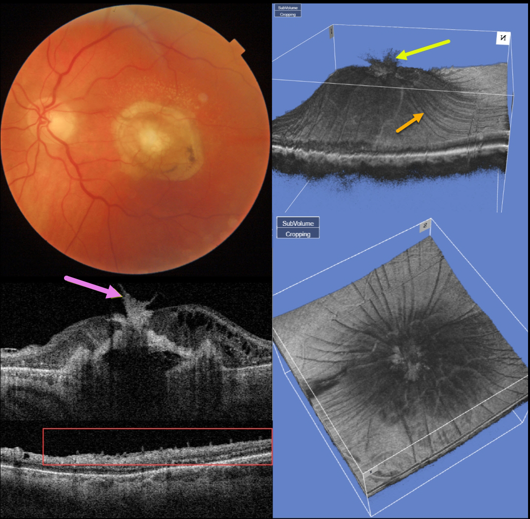
This 77YO man presented counting fingers vision OS with what seems at first to be an uneventful fibrotic disciform scar from neovascular AMD. But multimodal imaging shows several interesting findings.
Swept-source OCT through the macular center shows that the central scar extends through the full-thickness retina and becomes incorporated with the attached vitreous.
A 3D macular reconstruction viewed from above also shows how scar contraction is creating radiating retinal folds.
These folds on a B-scan taken through the peripheral macula give the appearance of numerous needle-like projections emanating from the inner retina.
The needle-like projections, created by imaging the radiating inner retinal folds perpendicular to their orientation, mimic findings felt to be pathognomonic for ocular amyloidosis (Kakuhara et al, Ophthalmology Retina 2021;5:1005-1008).



