

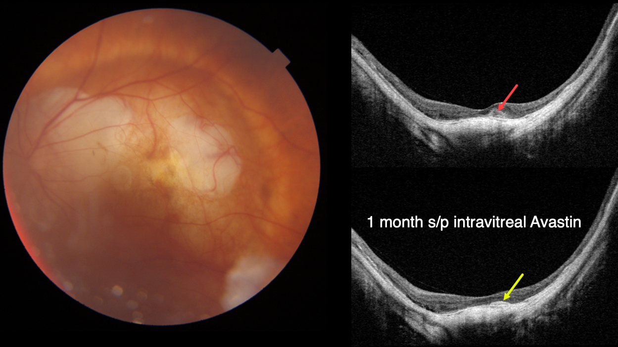
This 92YO female presented with new vision loss of 20/200 in her left eye. She has a history of high myopia, with hand motion vision OD due to a macular staphyloma. Baseline vision in her left eye is 20/40.
Fundus photography shows peripapillary and mostly superior macular atrophy.
Swept-source OCT shows a posterior staphyloma with a dome appearance to the central macula. A new small hyperreflective macular neovascularization (MNV) is noted.
One month following an intravitreal Avastin injection, vision improved to 20/40 with shrinkage of the MNV.
Learning Points:
In our experience, myopic MNV presents quite differently from those found in neovascular AMD.
Significant subretinal blood, subretinal fluid or intraretinal fluid are rarely seen. Often an active MNV will present as a new dot of subretinal blood or pigment.
Occasionally there will just be symptomatic vision loss or metamorphopsia without clinical or OCT findings.
Anti-VEGF therapy is effective, and these lesions can often be managed with PRN therapy.



