

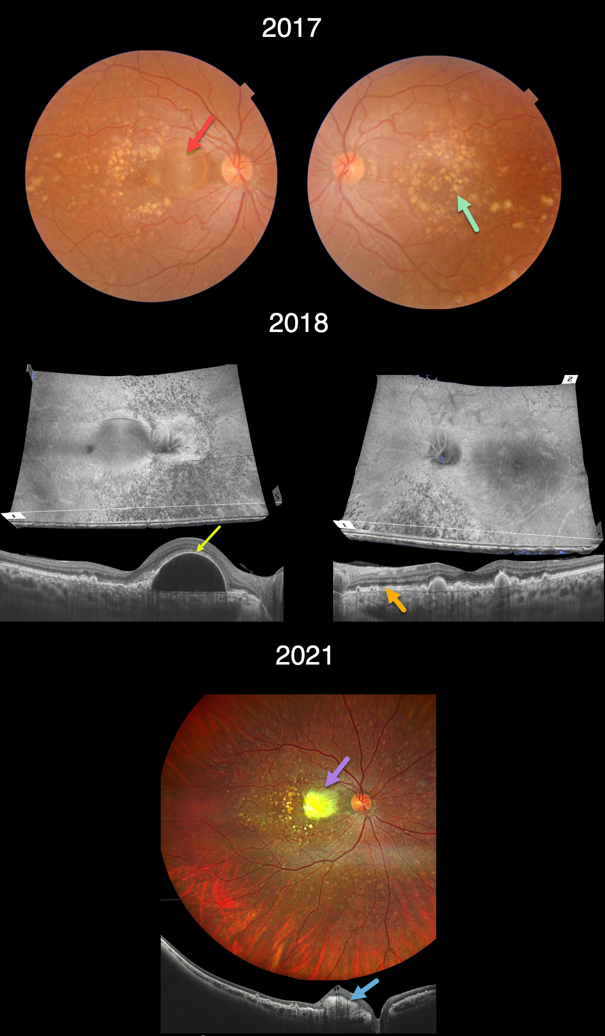
This 33YOF presented in 2017 with a recent history of vision loss in her right eye. She had no past ocular or medical history, nor family history of eye disease. Vision of 20/70 OD and 20/20 OS.
Triton color imaging shows extensive, mostly large drusen scattered throughout each macula, extending into the midperipheries. A large serous retinal pigment epithelial detachment (PED) is noted in the nasal right macula.
Triton swept-source OCT analysis in 2018 shows numerous unique perspectives. 3D reconstruction reveals extensive lumpy drusen extending throughout the posterior poles, especially outside each macula. B-scans show extensive variably elevated drusen. The large PED is evident in the nasal right macula. The retina external to the ellipsoid zone is thickened and hyperreflective.
Three years later vision decreased to 20/200 OD and was stable at 20/20 OS. The PED became clinically yellow and markedly hyperreflective on OCT (Canon Xephilio OCT-S1). The lesion failed to respond to anti-VEGF therapy.
Learning Points:
Doynes, also known as dominantly inherited radial basal laminar drusen or Malattia Leventinese, is a rare macular disorder caused by a mutation in the EFEMP1 gene on chromosome 2p16.
The EFEMP1 protein is a member of the fibulin family of extracellular matrix glycoproteins. The defective protein creates an abnormally thickened RPE basement membrane.
Centrally large, nodular,and confluent drusen are noted, along with a temporal radiating pattern of smaller cuticular drusen.
Later, there may be variable amounts of RPE hyperplasia and fibrous metaplasia, which is likely the cause of the vision loss in our patient’s right eye. Macular neovascularization may also develop.



