

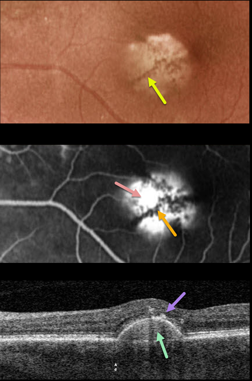
This 75YO female has been followed for several years with a stable, asymptomatic chronic retinal pigment epithelial detachment (PED) and 20/40 vision.
Optos color imaging shows a solid, variably depigmented, round PED in the inferonasal region of the left fovea. Radiating linear pigmentation is noted.
Fluorescein angiography shows hyperfluorescent pooling beneath the PED, with blockage from the overlying radiating pigment.
Swept-source OCT shows the PED with a hyperreflective core, overlying hyperreflective foci (representing the clinical radiating pigment), and a relatively thickened choroid for a patient of this age.
Learning Points:
Retinal pigment epithelial detachments are common in patients with AMD and are often associated with macular neovascularization. Non-vascularized PED is also seen as part of the pachychoroid spectrum.
The overlying pigment figure is pathognomonic for a chronic PED, and is seen even when these lesions flatten. Isolated avascular PEDs such as these do not require treatment.
Although our patient was in her mid-70s, there were no drusen and each fundus was otherwise normal. The chronicity of the PED, the lack of other signs of AMD, and the relatively thickened choroid all point to this being an avascular, pachychoroid PED.



