

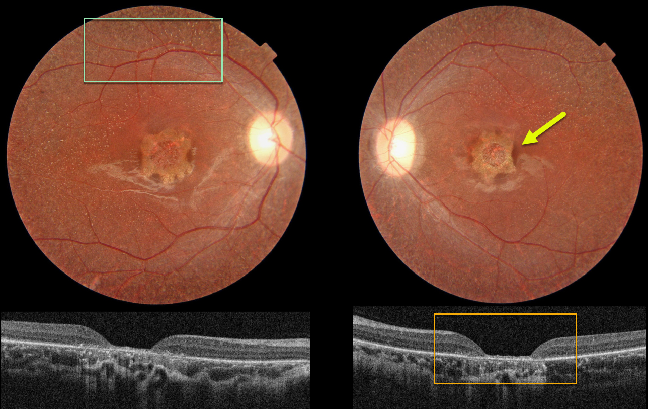
This 19YO female has stable 20/200 vision bilaterally with some form of inherited retinal disease (IRD).
Color photography shows a central one-disc-diameter area of scalloped atrophy with tiny white dots extending from the peripheral macula to beyond the arcades.
OCT shows central neurosensory retinal atrophy with a thin remaining rim of featureless tissue and underlying RPE loss.
Learning Points:
These findings do not fit neatly into any specific diagnosis. Stargardt/ABCA4 disease usually shows more pericentral than central atrophy, and usually involves just the outer retina. The subretinal flecks are much larger and yellow in appearance than the tiny whitish dots seen in our patient.
Fundus albipunctatus is a form of congenital stationary night blindness with somewhat similar white dots, but foveal atrophy and severe central vision loss are usually absent (though it may be associated with an RDH5 mutation).
Finally, retinitis punctata albescens presents with white dots similar to those in fundus albipunctatus, but it is a progressive disorder that can later develop more typical retinitis pigmentosa findings. Foveal atrophy is not a feature of this disease.
Fundus autofluorescence (FAF) is often helpful in differentiating these disorders. The lesions in ABCA4 disease are hyper-FAF. The global FAF signal is markedly diminished in fundus albipunctatus.
Unfortunately, our patient did not get FAF imaging. And of course, genetic testing would hopefully be diagnostic, but this was also not available.



