

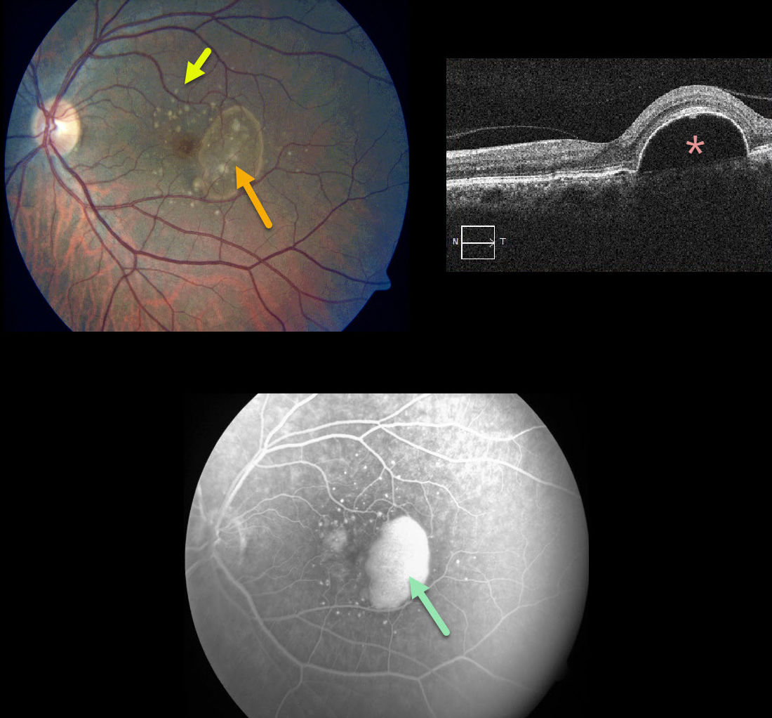
This patient presented with 20/25 vision in her left eye with significant Amsler grid changes. There is an irregular RPE detachment (PED) with variably sized drusen, and OCT confirms a large PED.
Fluorescein angiography shows a slightly uneven pattern of pooling beneath the PED.
We discussed starting treatment due to her symptoms, but for now, we have elected for close observation. There is a risk for an RPE tear with or without treatment
Learning Points:
PEDs associated with AMD are often associated with macular neovascularization (MNV).
Angiographic clues for MNV include a notched PED or uneven angiographic filling. Fluorescein angiography is probably still better than OCT angiography for detecting MNV.
Anti-VEGF therapy can effectively flatten these PEDs even when they are presumably avascular.



