

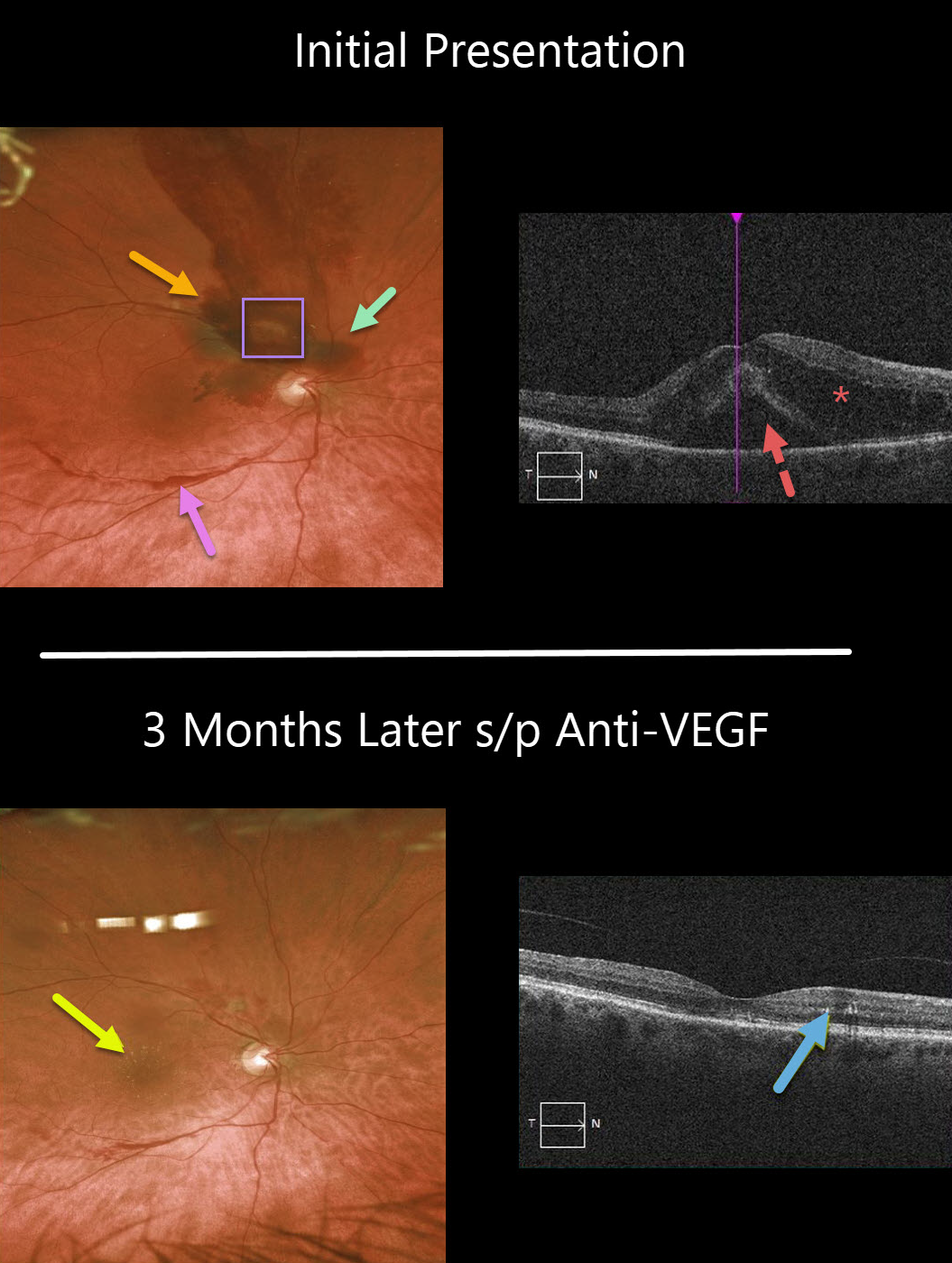
This 71YO female presented with counting-finger vision in her right eye. Clinically, there are multiple layers of blood, including preretinal, intraretinal, and subretinal. These multiple layers are virtually pathognomonic for a ruptured retinal arterial macroaneurysm (RAM).
Despite the causative RAM being just superotemporal to the optic nerve, significant intra- and subretinal fluid extends through the macular center, which is seen on OCT.
Following three monthly anti-VEGF injections, the fluid resolved completely, and vision returned to 20/30. Some residual precipitated lipid is seen in Henle’s layer as a faint lipid star in the color image and as hyperreflective dots in the outer plexiform layer.
Learning Points:
A retinal arterial macroaneurysm (RAM) is thought to result from a weakened arterial wall due to arteriosclerosis.
A RAM appears as a yellow-white dilated lesion along a retinal artery within the first 3 bifurcations from the optic nerve.
There are 2 distinct presentations, hemorrhagic or exudative. The hemorrhagic type often presents as a pathognomonic multilayer macular hemorrhage, with preretinal, intraretinal, and subretinal blood.



