

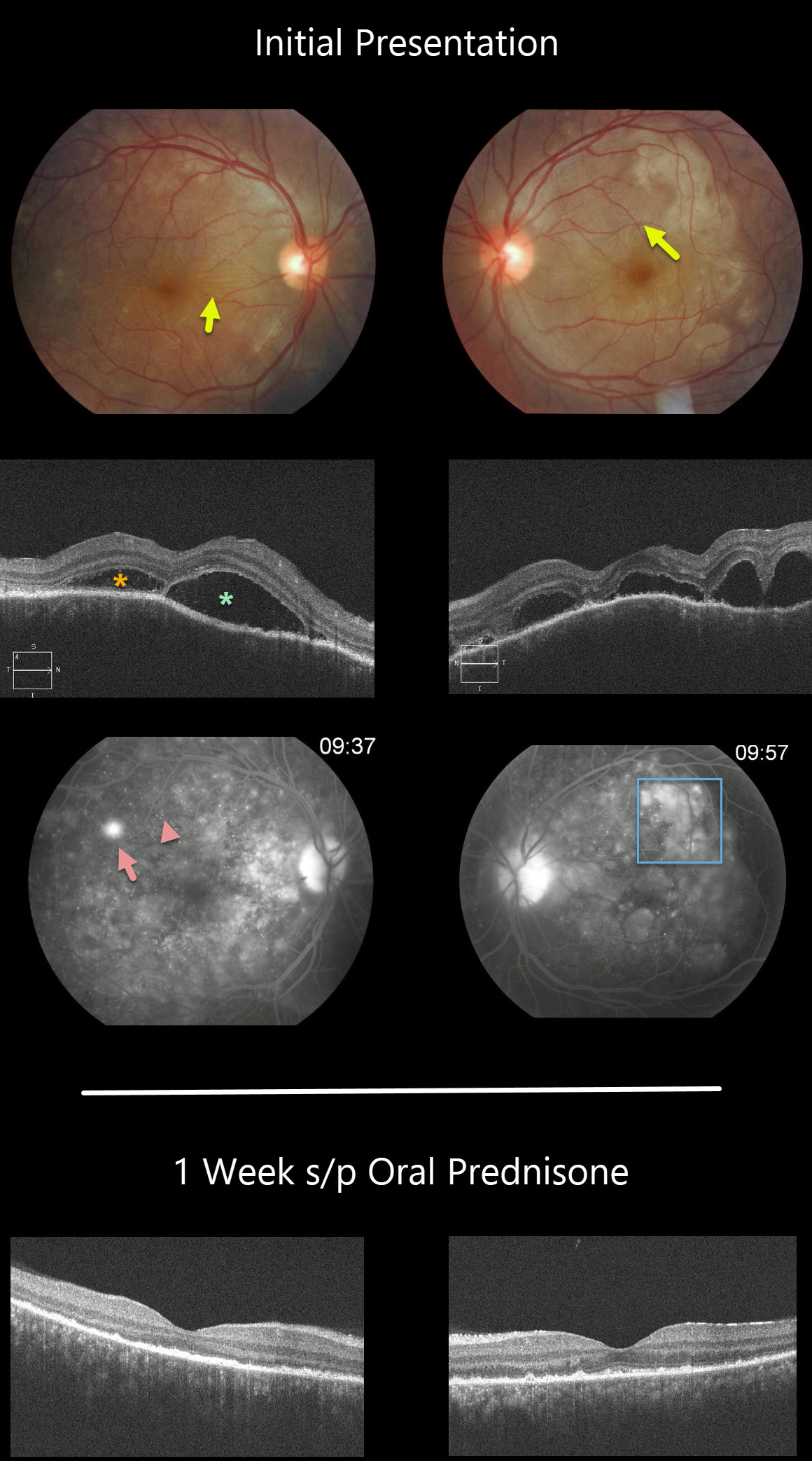
This patient has classic posterior segment findings of Harada disease, including multifocal exudative serous retinal detachments.
Choroidal inflammation produces a markedly thickened choroid with overlying chorioretinal folds (seen in both eyes as variably radiating outer retinal folds).
The OCT scans show multifocal serous detachments with bacillary detachments. Fluorescein angiography shows multiple subretinal leaks with pooling of fluorescein within the various subretinal and bacillary spaces.
The patient started 60mg of oral prednisone daily. One week later, there was a dramatic improvement, with complete resolution of all intra- and subretinal fluid and normalization of choroidal thickness.
Learning Points:
Harada disease is a bilateral granulomatous panuveitis. These patients often need extended treatment with oral prednisone. Most can be successfully weaned off of steroids, although chronic treatment is sometimes necessary.
Choroidal thickening is a good biomarker for disease activity and thins with controlled disease activity.
Remember that in Harada disease, there are only ocular findings. Vogt-Koyanagi-Harada disease occurs when there are associated auditory, integumentary or neurologic symptoms.



