

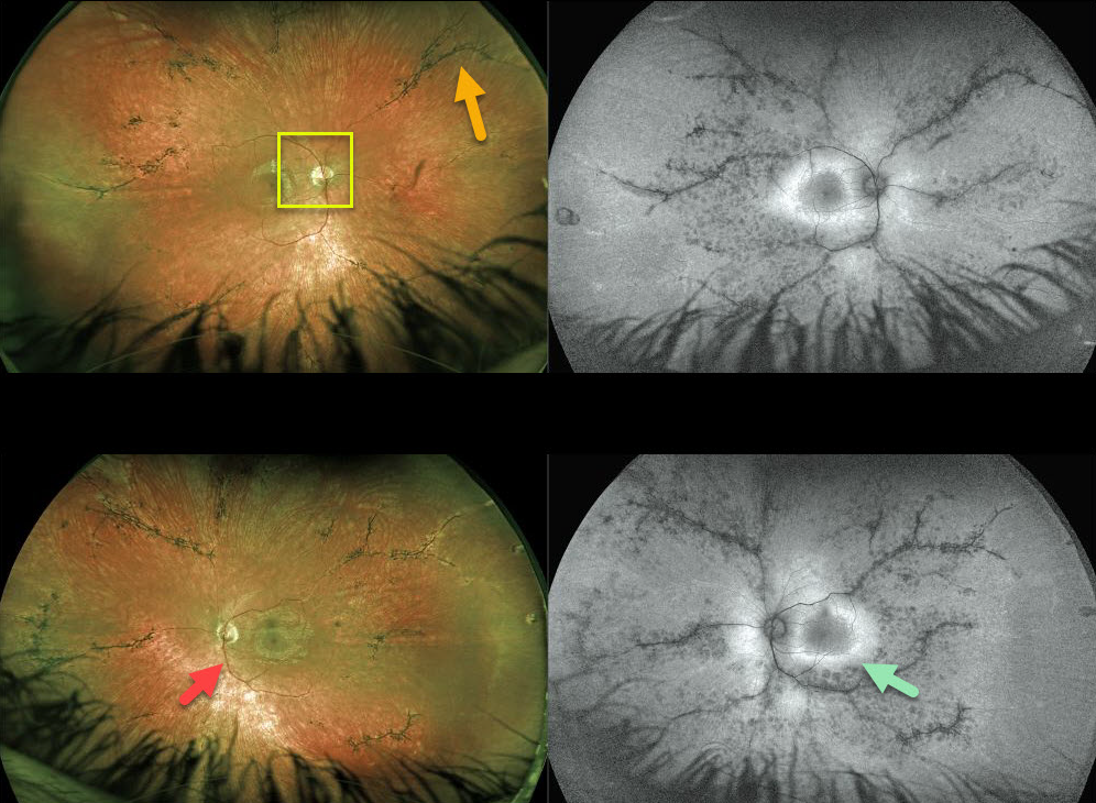
This 16YO patient with pigmented paravenous chorioretinal atrophy has excellent central vision of 20/20 OU but with significant nyctalopia.
Posterior segment findings include attenuated retinal vessels, bone spicules (intraretinal migration of the RPE along retinal capillaries), and optic disc pallor.
The bone spicules in this patient are primary perivascular, indicating the likely diagnosis of pigmented paravenous chorioretinal atrophy (PPCRA).
Fundus autofluorescence for this patient shows a ring of hyperautofluorescence around each central macula. This ring identifies at-risk retina for future degeneration and can help monitor the progression of this disease.
Learning Points:
Pigmented paravenous chorioretinal atrophy (PPCRA) is a rare condition characterized by paravenous pigment clumps with peripapillary and radial zones of RPE atrophy. It is bilateral, often asymmetric, with various causes, including genetic and pseudo-PPCRA (inflammatory, infectious, or unknown). Patients tend to have some symptoms, usually nyctalopia, but most retain good vision, especially those without macular involvement. The underlying pathophysiology is likely a primary choroidopathy that causes overlying RPE and retinal changes. The chorioretinal changes are best visualized with fundus autofluorescence.



