

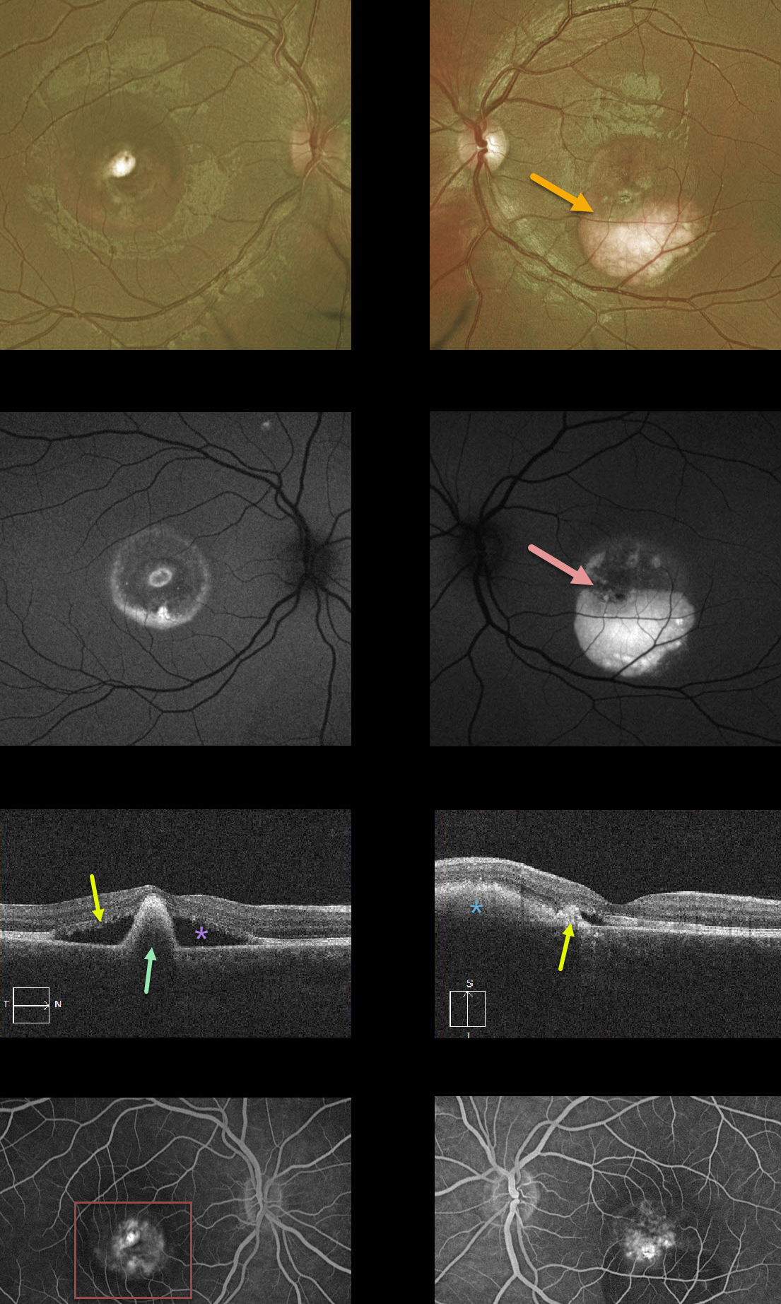
This patient has Best’s disease and has progressed to the more atrophic, ‘scrambled egg’ appearance. The fundus photos show asymmetric layering of the residual yellow-white subretinal vitelliform material.
This material is highly autofluorescent and can also appear as hyperreflective on OCT scans. The right eye has a large subfoveal pigment clump with surrounding non-exudative subretinal fluid. The left eye has a smaller subfoveal scar with a flattened vitelliform lesion without subretinal fluid. Fluorescein angiography (FA) shows staining without active leakage.
Learning Points:
Best vitelliform macular dystrophy (Best’s disease) is associated with a mutation in the BEST1 gene. This inherited disorder usually presents in childhood or early adulthood with a bilateral foveal “egg-yolk” appearance.



