

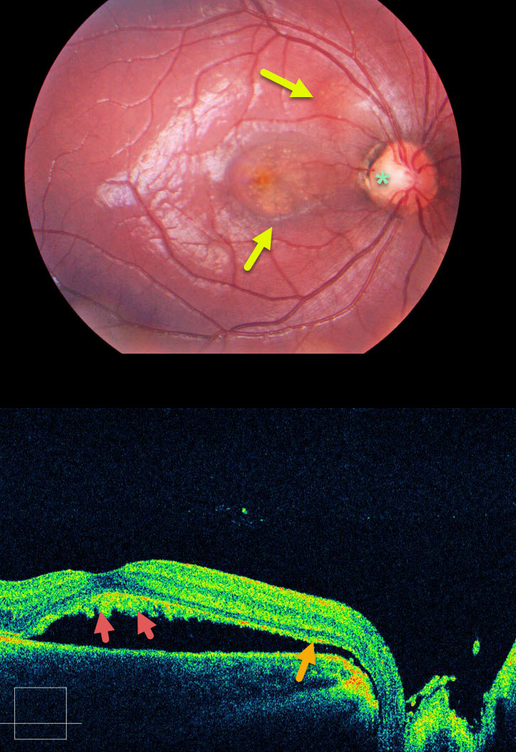
This patient has optic pit maculopathy, in which areas of subretinal fluid can be traced back to the optic nerve pit via an abnormal communication between the subretinal and subarachnoid spaces.
Shed photoreceptor outer segments accumulate on the back surface of the detached retina since the RPE cells can’t perform their normal function of phagocytosis. This shaggy vitelliform material gives the central macula a yellow appearance.
Learning Points:
Optic pits are congenital defects caused by the incomplete closure of the fetal fissure during embryogenesis. Macular detachment and schisis occur in up to 30-40% of eyes.
When symptomatic, a myriad of surgical procedures have been described. As with any problem with too many solutions, there is usually no best option.
A recent meta-analysis concluded that just a plain, vanilla vitrectomy is probably as successful as anything else (Zheng et al, Ophthalmology Retina 2020;4:389-299).



