

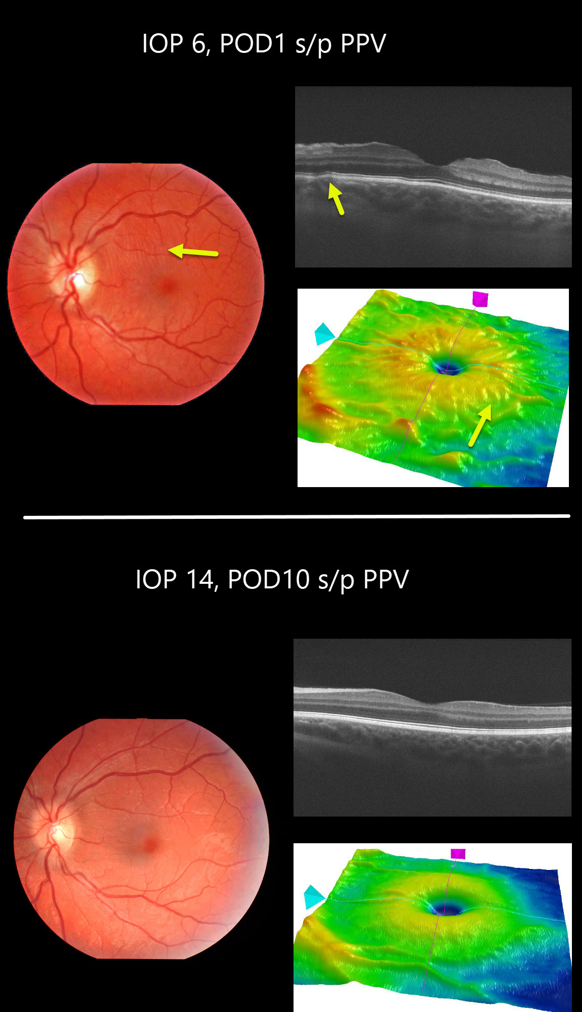
This patient developed hypotony maculopathy one day following vitrectomy for vitreous floaters. Intraocular pressure was 6mmHg and all sclerotomy sites appeared tight. The retinal vessels are somewhat engorged and dilated.
Chorioretinal folds are seen in the fundus photo, OCT B-scan and thickness map.
These findings completely resolved 9 days later when his IOP recovered spontaneously to 14mmHg.
Learning Points:
Hypotony maculopathy is characterized by low IOP and chorioretinal folds, most commonly following glaucoma filtration surgery. Optic nerve edema can also be seen due to decreased axoplasmic transport.



