

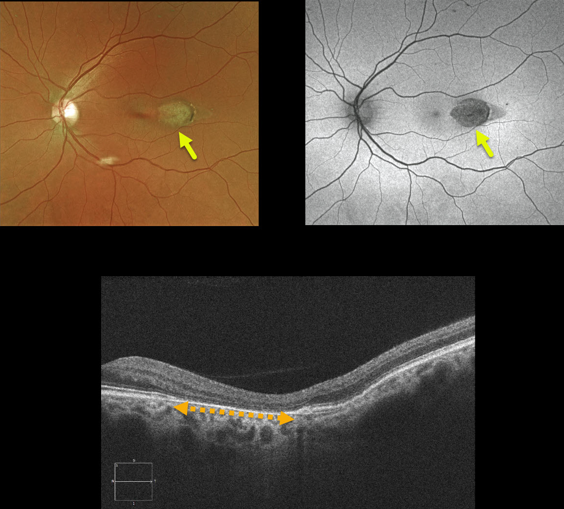
This patient has torpedo maculopathy, originally described by Dr. Gass as a solitary hypopigmented nevus of the RPE.
These benign, unilateral lesions are teardrop-shaped and almost always located in the horizontal meridian of the temporal macula. OCT shows attenuation of the RPE and outer retinal layers.
Vision is almost always normal.
Learning Points
These torpedo lesions are congenital with no systemic associations. Congenital hypertrophy of the RPE and Gardner’s syndrome-associated RPE lesions can mimic torpedo maculopathy.
See Villegas et al, Ophthalmic Surg Lasers Imaging Retina 2014;45:222-226, for a great discussion on distinguishing these RPE lesions.



