

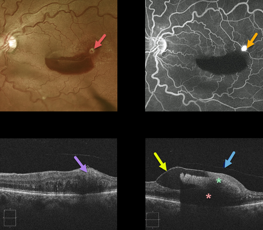
This retinal arterial macroaneurysm (RAM) appears as a yellow-white inner retinal lesion along the superotemporal edge of a macular hemorrhage. Angiography confirms the presence of the hyperfluorescent RAM.
An OCT line scan through the RAM shows its location in the inner retina.
OCT scanning shows that the blood is almost all above the retina and below the detached internal limiting membrane. The detached hyaloid can be seen anterior to the ILM. The dense sub-ILM blood blocks almost all retinal and subretinal detail more posteriorly.
Learning Points:
A retinal arterial macroaneurysm (RAM) is thought to result from a weakened arterial wall due to arteriosclerosis.
A RAM appears as a yellow-white dilated lesion along a retinal artery within the first 3 bifurcations from the optic nerve.
There are 2 distinct presentations, hemorrhagic or exudative. The hemorrhagic type often presents as a pathognomonic multilayer macular hemorrhage, with preretinal, intraretinal, and subretinal blood.



