

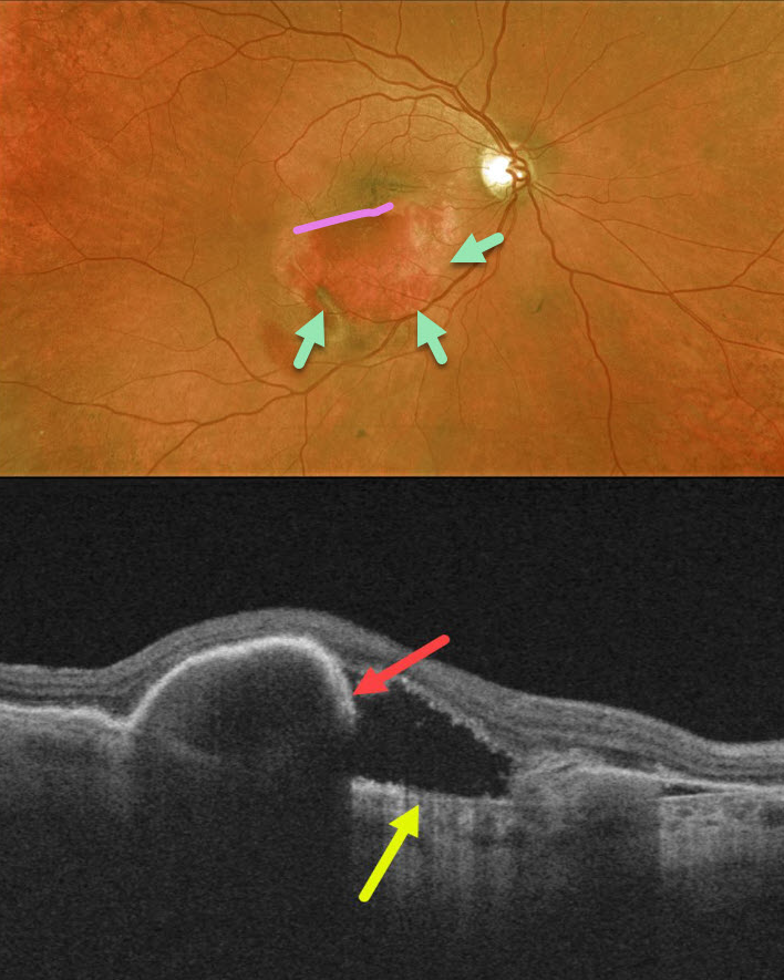
Our patient had a retinal pigment epithelial (RPE) tear 2 weeks following an injection for wet age-related macular degeneration. The edges of the bare choroid and the edge of the retracted RPE can be visualized.
OCT shows bare Bruch’s membrane and the retracted, torn RPE.
Learning Points:
A tear of the RPE is a fairly common finding in RPE detachment (PED) associated with type 1 macular neovascularization (MNV), which is located below the RPE.
The neovascularization can spontaneously contract or involute following treatment. It then puts traction on the overlying RPE, which can then rip at the opposite edge of the PED. Like a cheap window blind, the torn RPE retracts and scrolls towards the MNV. This leaves a depigmented region where the RPE was and is now absent, along with a pigmented, relatively straight subretinal lesion extending from the scrolled RPE edge.



