

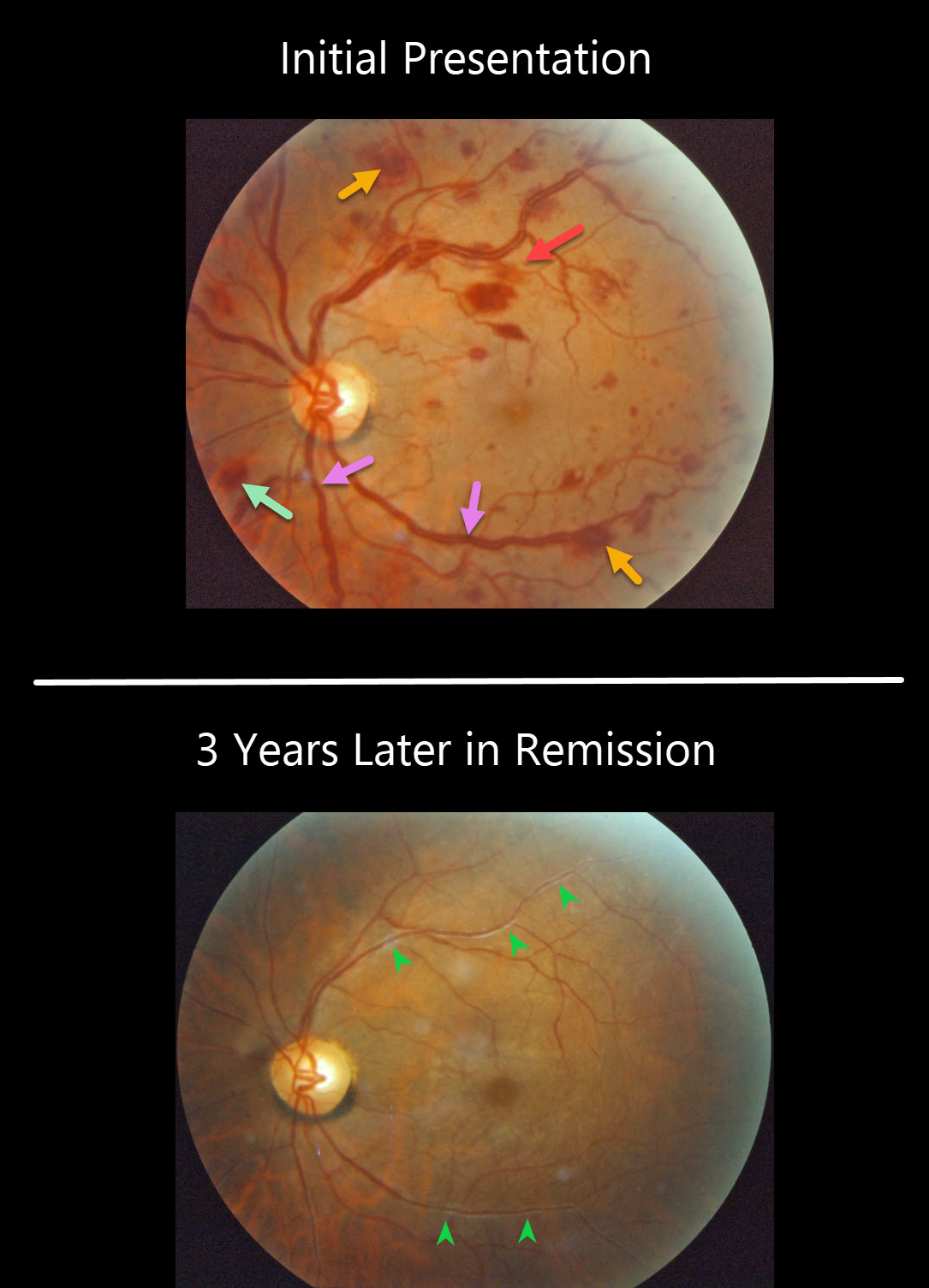
This patient presented with bilateral multiple flame-shaped and white-centered retinal hemorrhages. There is also at least one deeper, round outer plexiform hemorrhage just inferonasal to the nerve. The retinal veins are somewhat dilated and tortuous, consistent with hyperviscosity. This patient was subsequently diagnosed with multiple myeloma.
Three years later, the patient was in remission after receiving treatment. Although the vascular caliber has normalized, vascular sheathing remained due to prior damage to the vessel walls.
Learning Points:
White-centered hemorrhages or “roth spots” can be seen in many disorders, including leukemia, hyperviscosity, anemia, diabetes, and hypertension. The white material may represent a variety of causes, including fibrin.
The term “Roth spot” was originally named after Dr. Moritz Roth, who, in 1872, noted white-centered hemorrhages in patients with bacterial endocarditis. He felt that these white spots were septic emboli that originated from an infected cardiac valve.
This term should thus be reserved only for patients with bacterial endocarditis. See Duane et al Ophthalmology 1980;87;66-69.



