

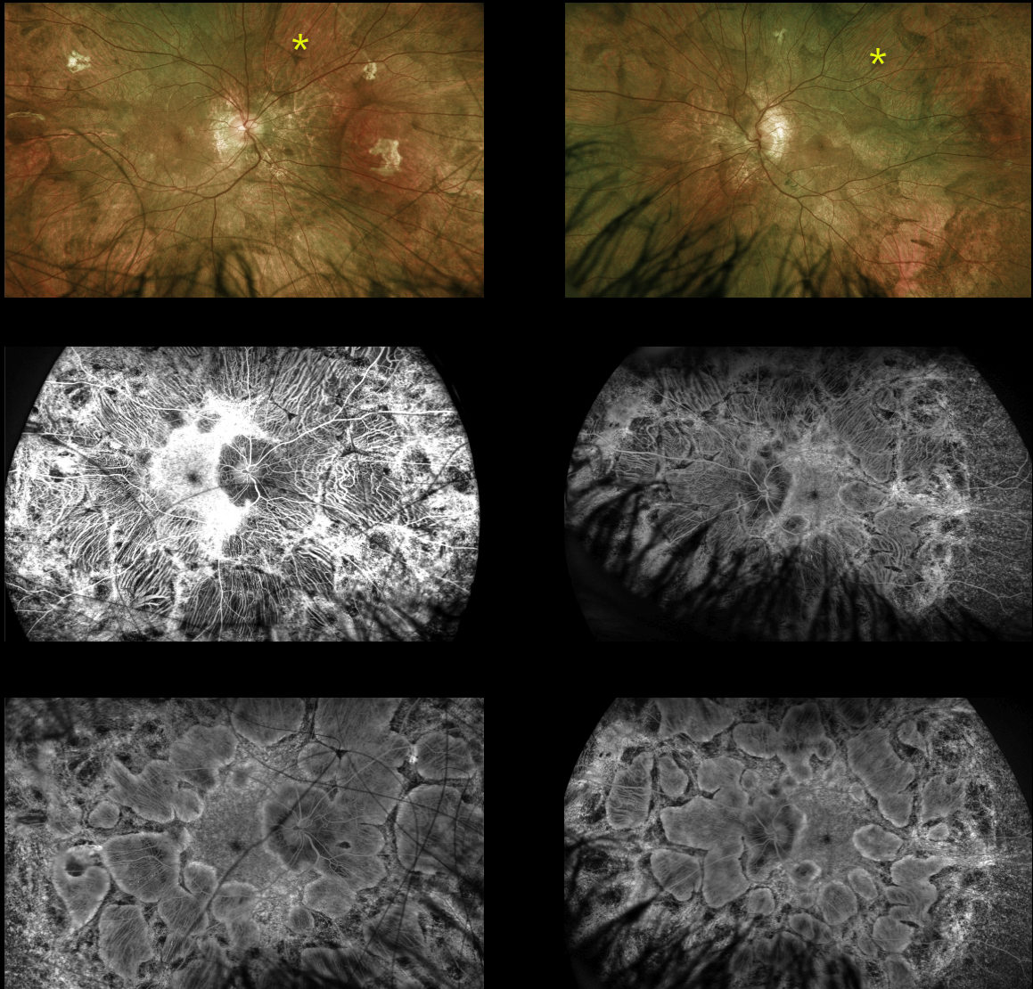
This 14YO male is surprisingly asymptomatic with visual acuity of 20/30 OD and 20/40 OS. His retinal findings were found on a routine exam.
Optos imaging shows large symmetrically bilateral scalloped areas of chorioretinal atrophy, which spare the central maculas. On fundus fluorescein angiography, the early phase (middle) shows exquisite detail of the choroidal vortex system and late phase (bottom) shows staining of the scalloped areas of atrophy.
The patient is adopted and is unaware of any family history of inherited retinal disease. Subsequent genetic testing revealed a pathogenic hemizygous CHM variant on the X chromosome.
Learning Points:
Choroideremia is an X-linked recessive chorioretinal dystrophy caused by a mutation in the CHM gene located on the long arm of the X chromosome. Sons of female carriers have a 50% chance of developing choroideremia, and daughters have a 50% chance of becoming carriers.
Some female carriers can still develop clinical disease due to irregular inactivation of the X-chromosome (see Jauregui et al, AJO 2019;207:77-86).
The onset of night blindness usually begins between the ages of 10 and 30, followed by peripheral visual field loss. Zones of patchy RPE and chorioretinal atrophy initially appear in the mid-periphery, gradually spreading anteriorly and posteriorly.
The choroidal vortex system is relatively resistant to most disorders, including choroideremia. This system is beautifully imaged angiographically in our patient since the RPE, which normally blocks the underlying choroid, is atrophic and no longer blocks these vessels.
Eventually, the patient is left with a small central island of vision, narrowed retinal vessels and optic atrophy.
For a great choroideremia review article, see Pennesi et al, Retina 2019;39:2019-2069.



